h1.qusth1 { display: none !important; }

QUESTION OF THE WEEK

Dr. Donovan's Articles

QUESTION OF HAIR BLOGS

Filtering by Category: Cicatricial


Compounding of Follicles

Compounding of Follicles

compound follicles.png

Compound follicles are hair follicle units that have more than 6 hairs coming out of a single pore. The tendency to form compound hair follicles is sometimes a feature of so called neutrophil mediated scarring alopecias. Folliculitis decalvans (shown here) frequently shows compounding of hairs. 


This article was written by Dr. Jeff Donovan, a Canadian and US board certified dermatologist specializing exclusively in hair loss.
Share This
No Comments

Perifollicular erythema in FFA

Redness around hair follicles: Perifollicular eythema

Frontal fibrosing alopecia or "FFA" is a scarring alopecia (scarring hair loss condition) that most commonly develops in women 45-65. It causes permanent hair loss. The cause is not know although a mix of immune based mechanisms and hormonal mechanics are likely to contribute. The condition can be asymptomatic - and many patients have no itching, burning or pain.


FFA Disease Activity: How do we know it's' active or not

PFE

How do we know whether a given patient's FFA is active? Undoubtedly, the absolute best way is with a photo. If a patient's photo changes over a period of monitoring (6 months or 12 months) the FFA is active by definition.

However, photographs don't capture subtle changes in activity. To accomplish this examination by "dermoscopy" is helpful. In this photo, slight redness around the hairs can be seen. We call this "perifollicular" erythema. (note peri means "around"). In 2013, Spanish researchers Toledo- Pastrana and colleagues published an article in the International Journal of Trichology examining dematoscopic features of FFA. Of 79 patients examined, 66 % showed perifollicular erythema. In patients with active disease, perifollicular erythema was present in 95 % of patients.


Conclusion


Perifollicular erythema is an important sign to look for in FFA. It indicates disease activity and a high likelihood of further hair loss in the patient.


Reference


Toledo- Pastrana et al. Perifollicular erythema as a trichoscopy sign of progression in frontal fibrosing alopecia. Int J Trichoscopy 2013; 5: 151-3.


This article was written by Dr. Jeff Donovan, a Canadian and US board certified dermatologist specializing exclusively in hair loss.
Share This
No Comments

Dissecting Cellulitis: Early Features

Early Stages of DSC

Dissecting Cellulitis (DSC) is a relatively rare scarring alopecia. Men are affected much more than women and affected patients are frequently young males in their 20s.

dsc2

The condition beings with discharge of pus and sometimes blood from the scalp. It can also be quite itchy. The beard, underarm (axilla) and groin can be affected with similar discharge. A key feature of diagnosis is the presence of sinus tracts or "tunnels" underneath the skin. Small thin vellus hairs are also seen in the affect area.

The primary treatment of DSC is isotretinoin although antibiotics, zinc, dapsone, colchicine, and TNF inhibitors can be used.


This article was written by Dr. Jeff Donovan, a Canadian and US board certified dermatologist specializing exclusively in hair loss.
Share This
No Comments

Pseudopelade of Brocq

ppb2.png

Pseudopelade of Brocq

Pseudopelade of Brocq (PPB) is a scarring alopecia. It causes permanent hair loss. The cause is unknown.

In contrast to lichen planopilaris, there is little to no scale around hair follicles. The areas may be pink-colored when active. Treatment is similar to lichen planopilaris including use of topical steroids, steroid injections, topical calcineurin inhibitors, oral methotrexate, oral doxycycline, oral hydroxychloroquine and others.


This article was written by Dr. Jeff Donovan, a Canadian and US board certified dermatologist specializing exclusively in hair loss.
Share This
No Comments

Pseudopelade of Brocq

Pseudopelade of Brocq

ppb.png

Pseudopelade of Brocq (PPB) is a scarring alopecia that affects both women and men. The central scalp is often affected first. This condition causes permanent hair loss - hair does not regrow. The goal of treatment is to stop further loss. The cause is unknown.

The areas of hair loss are usually pale colored in those with PPB although they may be slightly pink. When one feels these areas with a finger it is usually obvious that the area dips down below the level of the skin. We call this phenomenon "atrophy". The condition is frequently misdiagnosed as lichen planopilaris (another scarring hair loss condition). The two conditions are similar but lichen planopilaris has more redness and scaling than PPB and is generally more responsive to treatment. Treatment of PPB is similar to LPP and includes topical steroids, topical calcineurin inhibitors, doxycycline, hydroxychloroquine, steroid injections and similar anti-lymphocytic agents.


This article was written by Dr. Jeff Donovan, a Canadian and US board certified dermatologist specializing exclusively in hair loss.
Share This
No Comments

Biologic Agents and LPP: What do they teach us?

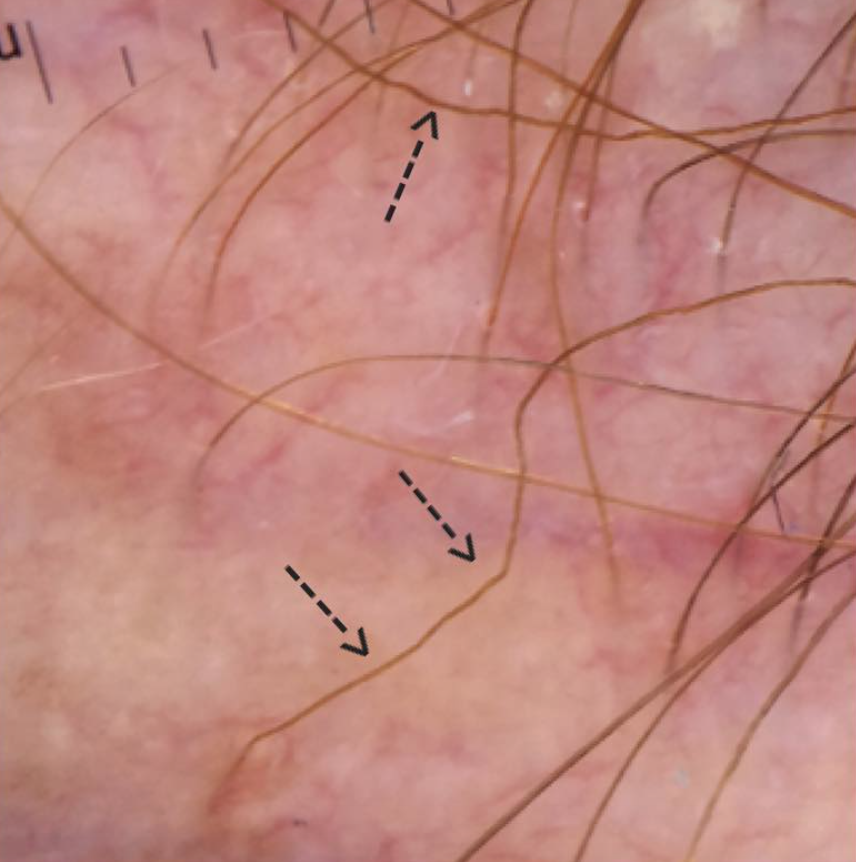
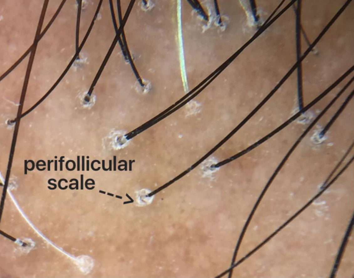
Biologic Agents in Lymphocytic Scarring Alopecia

The field of dermatology has changed dramatically with the introduction of a variety of monoclonal antibodies, fusion proteins and growth factors to the list of treatment options. These agents are used to treat a wide range of disorders including psoriasis, hidradenitis suppurativa, atopic dermatitis, pyoderma gangrenosum, and various blistering diseases. This is by no means the full list.

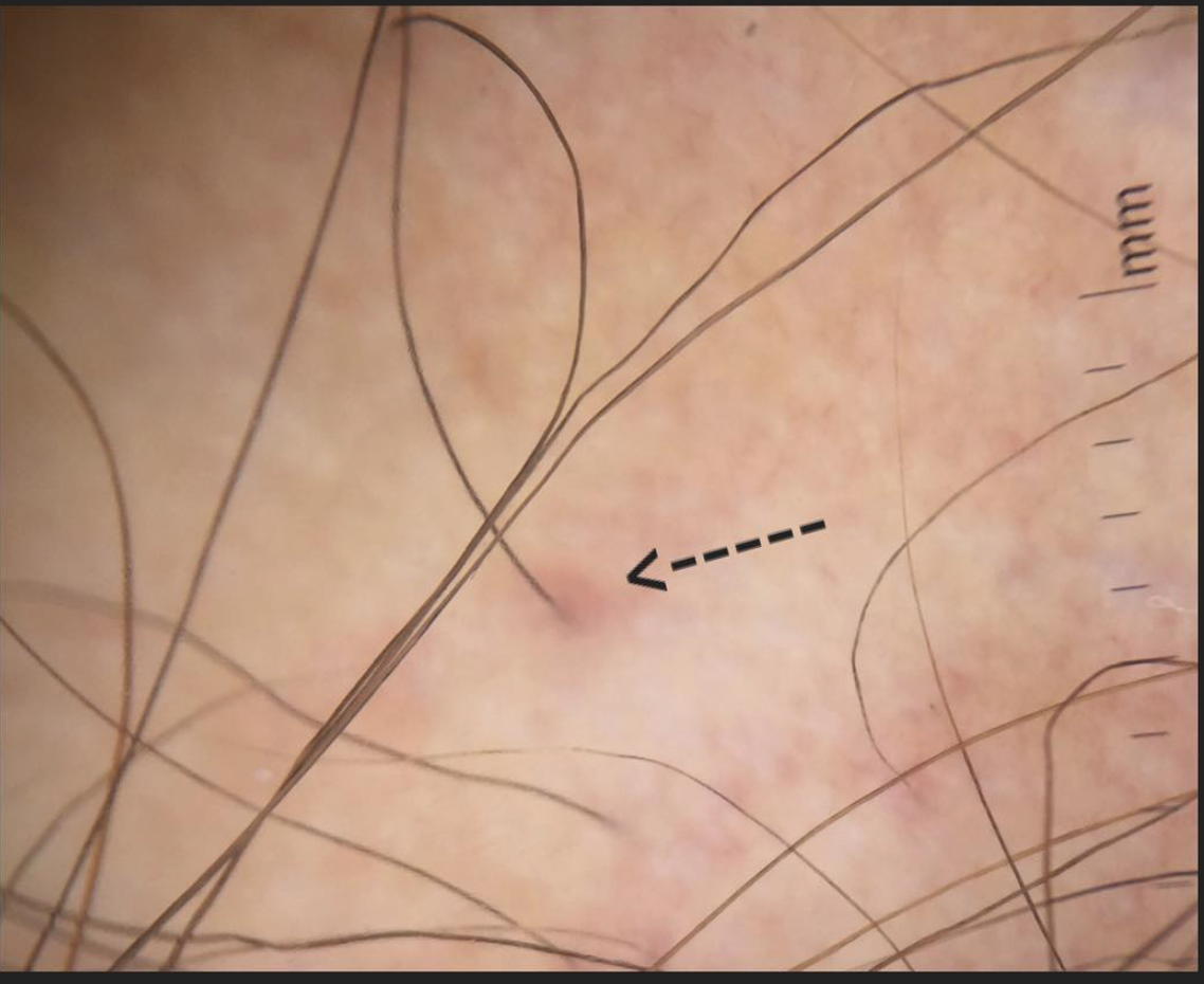
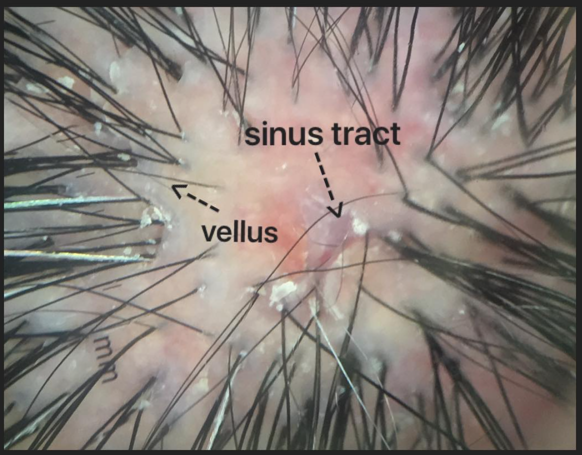
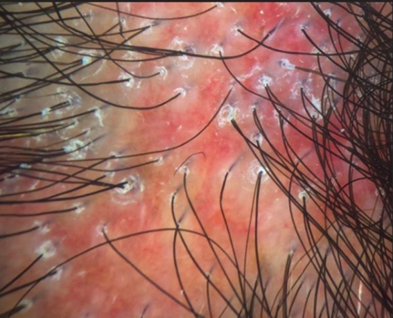
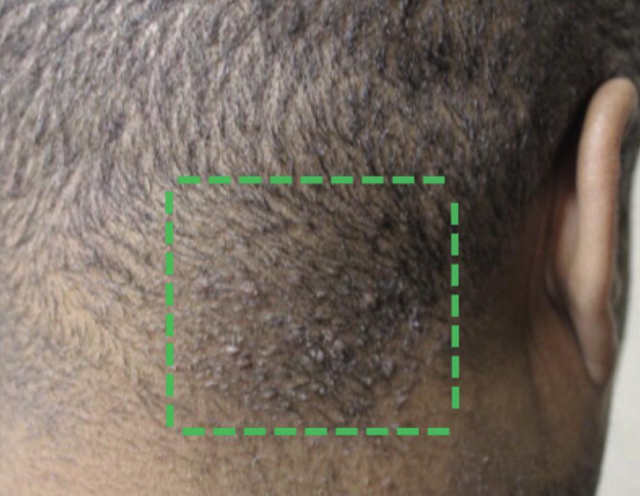
The use of these agents in lymphocytic scarring alopecias like lichen planopilaris has been studied to a very limited degree. However their benefits are not entirely clear and many agents may actually worsen the disease. Here, we very briefly review some of the existing literature about the use of various biologic agents in treating LPP, including the monoclonal antibodies, fusion proteins and growth factors.


CATEGORY 1: Monoclonal Antibodies

Monoclonal antibodies are antibodies that target specific cell-surface receptors. These antibodies may be chimeric, humanized or human depending on how much murine and human proteins they contain. 


a) Anti-TNF Antibodies: Adalimumab, Infliximab, Certolizumab, Golimumab

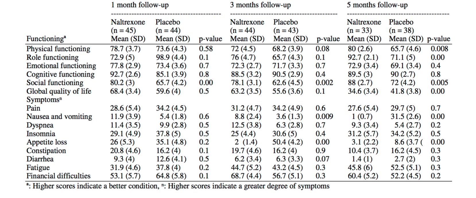
TNF-α antagonists have been associated with paradoxical psoriasiform, lichenoid, eczematous, granulomatous, and acneiform eruptions.  Lichenoid reactions specifically are uncommon but are an emerging cutaneous adverse effect.   In 2010, Fernandez-Torres reported the development of lichen planopilaris in with infliximab treatment. The patient in the case was a 37-year-old man with recalcitrant plaque psoriasis who was being treated with infliximab at a dosage of 5 mg/kg every 8 weeks. However, 11 months into treatment he presented with follicular keratotic papulo-pustules, perifollicular erythema, and scaling, with progressive hair loss of the frontal and parietal regions of the scalp and eyebrows. In 2016, Jayasekera and colleagues reported the development of lichen planopilaris in a 12 year old girl treated with adalimumab for oliogarthritis. Her lesions cleared with discontinuation of the clobetasol.


b) Anti-CD20: Rituximab

Rituximab is a chimeric monoclonal antibody against the protein CD20, which is primarily found on the surface of immune system B cells. In 2011, Erras and colleagues published a report in the European Journal of Dermatology of a patient with juvenile arthritis treated with rituximab who experienced complete resolution of co-existening lichen planopilaris.


c) Anti-IL-12 and anti-IL-23 monoclonal antibody: Ustekinumab

In 2015, Weber published a report of a patient with LPP treated with ustekinumab over a 10 months period without evidence of any benefit 


d) Anti-LFA1: Efalizumab

To the best of my knowledge, there are no reports of lichen planopilaris with this group of agents no evidence of a treatment benefit with these agents. 


e) Anti-CD2 antibody: Siplizumab

To the best of my knowledge, there are no reports of lichen planopilaris with this group of agents no evidence of a treatment benefit with these agents. 


f) Anti-CD4 antibody: Orthoclone (OKTcdr4a)

To the best of my knowledge, there are no reports of lichen planopilaris with this group of agents no evidence of a treatment benefit with these agents. 


g) Anti-CD25 antibodies: Basiliximab, Daclizumab

To the best of my knowledge, there are no reports of lichen planopilaris with this group of agents no evidence of a treatment benefit with these agents. 


h) Anti-CD80r: Galiximab (IDEC 114)

To the best of my knowledge, there are no reports of lichen planopilaris with this group of agents no evidence of a treatment benefit with these agents. 


i) Anti-IgE: Omalizumab

To the best of my knowledge, there are no reports of lichen planopilaris with this group of agents no evidence of a treatment benefit with these agents. 


CATEGORY 2: Fusion Antibody Proteins

Fusion proteins, also known as 'chimeric proteins', are proteins which are created by the fusion of the receptor domain of a human protein with the constant region of human IgG. The resultant fusion protein then attaches (binds) specifically to a ligand or co-receptor. Recombinant fusion proteins have also been produced by combining human proteins with bacterial toxins. The fusion proteins most commonly used in dermatology are Etanercept, Alefacept, Abatacept, and Denileukin Diftitox. Of these four agents, only Etanercept has been studied in relationship to lichen planopilaris.

a) Etanercept

In 2009, Abbasi and colleagues reported the development of lichen planopilaris in a patient with psoriasis who was treated with etanercept.  Just the year before, Garovich reported a case of LPP with etanercept in the British Journal of Dermatology. 


b) Alefacept

To the best of my knowledge, there are no reports of lichen planopilaris with this group of agents no evidence of a treatment benefit with these agents. 


c) Abatacept

To the best of my knowledge, there are no reports of lichen planopilaris with this group of agents no evidence of a treatment benefit with these agents. 


d) Denileukin Diftitox

To the best of my knowledge, there are no reports of lichen planopilaris with this group of agents no evidence of a treatment benefit with these agents. 


CATEGORY 3: Recombinant human cytokines and growth factors

This category consists of water soluble non-immunoglobulin proteins and glycoproteins produced by a wide variety of cells. A variety of immune stimuli lead to their production and release.   Recombinant cytokines or cytokine antagonists are manufactured by recombinant DNA technology have been used as immunomodulators for malignant and inflammatory skin conditions.  The principal recombinant cytokines used in dermatology are interferon α (IFNα), Interferon γ (IFNγ), Interleukin 1 Receptor antagonist (IL1Ra), Interleukin 2 (IL-2), Interleukin 4 (rhIL-4), Interleukin 10 (rhIL-10), Interleukin 11 (rhIL-11), Granulocyte macrophage colony stimulating factor (GM-CSF), Platelet derived growth factor (PDGF). 

To the best of my knowledge, there are no reports of lichen planopilaris with this group of agents no evidence of a treatment benefit with these agents. 


REFERENCE

Abbasi NR, et al. Lichen planopilaris noted during etanercept therapy in a child with severe psoriasis. Pediatr Dermatol. 2009

Fernández-Torres R, et al. Infliximab-induced lichen planopilaris. Ann Pharmacother. 2010. 

Garcovich S, et al. Onset of lichen planopilaris during treatment with etanercept. Br J Dermatol. 2008

Jayasekera PS, et al. Case Report of Lichen Planopilaris Occurring in a Pediatric Patient Receiving a Tumor Necrosis Factor α Inhibitor and a Review of the Literature. Pediatr Dermatol. 2016 Mar-Apr.

McCarty M, et al. Lichenoid Reactions in Association with Tumor Necrosis Factor Alpha Inhibitors: A Review of the Literature and Addition of a Fourth Lichenoid Reaction. J Clin Aesthet Dermatol. 2015Erras S, et al. Rapid and complete resolution of lichen planopilaris in juvenile chronic arthritis treated with rituximab. Eur J Dermatol. 2011 Jan-Feb.

Webster G. Failure of lichen planopilaris to respond to ustekinumab.  Dermatol Online J. 2015.


This article was written by Dr. Jeff Donovan, a Canadian and US board certified dermatologist specializing exclusively in hair loss.
Share This
No Comments

What are the side effects of Low Dose Naltrexone (LDN)?

Low dose naltrexone: What side effects are possible?

Low dose naltrexone is increasingly studied in the field of hair loss, mainly in the areas of scarring alopecia and alopecia areata. To date, there remains only limited evidence that these drugs have a role. We continue to study them in our clinic. 

DOWNLOAD LDN HANDOUT


Side effects: What side effects are possible?

Dr. Donovan generally counsels patients about the top side effects including 


1. Difficultly sleeping.

This is usually just for the first week. If trouble sleeping go beyond this, one can reduce the dose to 3 mg or 1.5 mg


2. More vivid dreams.

This is seen in approximately 37 % of LDN users and can decrease over time.


3. Reduced need for thyroid medication.

Patients with autoimmune thyroid disease who take thyroid medications may want to start with a 1.5 mg dose and monitor their TSH every 2-4 weeks. This is to prevent a change from a hypothyroid/euthyroid state to a hyperthyroid state. Many patients with LDN require less thyroid supplementation while on LDN.

4.  Headaches.

Headaches have been noted to be increased compared to placebo. 


5. Anxiety (rare).

To date, there is not a consistent increase in headaches in the frequency of anxiety in LDN users compared to placebo.


6. Tachycardia (increased heart rate)

Although tachycardia is something we watch for, to date, there is not a consistent increase in headaches in the frequency of abnormal heart rhythms in LDN users compared to placebo in studies conducted to date.


7.   Rare - Fatigue, Loss of appetite, nausea, mood swings, mild disorientation


Side effects in the clinical studies

There are only a limited number of well conducted studies examining side effects of low dose naltrexone. By well conducted studies, one is specifically referring to studies that compared side effects of LDN to placebo. Here are some studies that guide our understanding of side effects of LDN


1.  Younger and colleagues, 2013. 

A study of 31 patients with fibromyalgia by Younger and colleagues showed that headaches and vivid dreams were by far the most important of the side effects of LDN compared to those using placebo.  Other side effects did not appear statistically different in LDN users vs placebo (at least based on the small numbers). 

SOURCE: Younger and colleagues. Arthritis Rheumatism 2013. 

SOURCE: Younger and colleagues. Arthritis Rheumatism 2013. 


2. Mischoulon et al, 2017

A randomized trial of 1 mg twice daily low dose naltrexone (LDN) was studied by Mischoulon and colleagues in 12 patients with depression. Their study was small but differences in side effects between the treatment (NTX) and placebo (PBO) groups were not appreciable. 

Mischoulon D, et al. J Affect Disord. 2017.

Mischoulon D, et al. J Affect Disord. 2017.


3. Laser Sharafaddinzadeh et al, 2010

In a randomized study study of 106 patients with multiple sclerosis, the main side effects were nausea, epigastric pain, mood alteration, mild irritability, headache, and joint pain.


4. Mohammad Ali Seifrabiei et al, 2008

A randomized study of low dose naltrexone in 89 hematologic patients showed that LDN was associated with better appetite, reduced nausea and vomiting compared to users of placebo. There were no differences in insomnia in this study. 

Mohammad Ali Seifrabiei et al. Am J Applied Sciences 2008

Mohammad Ali Seifrabiei et al. Am J Applied Sciences 2008


5. Smith et al, 2013

Small studies in children receiving low dose naltrexone for inflammation bowel disease (Crohn's disease) showed no differences in sleep, dreams, twitching, headaches, appetite, nausea, or double vision. 


References

Younger J, et al. Low-dose naltrexone for the treatment of fibromyalgia: findings of a small, randomized, double-blind, placebo-controlled, counterbalanced, crossover trial assessing daily pain levels. Randomized controlled trial. Arthritis Rheum. 2013.

Mischoulon D, et al. Randomized, proof-of-concept trial of low dose naltrexone for patients with breakthrough symptoms of major depressive disorder on antidepressants. Randomized controlled trial.  J Affect Disord. 2017.

Smith JP, et al. Safety and tolerability of low-dose naltrexone therapy in children with moderate to severe Crohn's disease: a pilot study. Randomized controlled trial.  J Clin Gastroenterol. 2013.

David Mischoulon et al. Randomized, proof-of-concept trial of low dose naltrexone for patients with breakthrough symptoms of major depressive disorder on antidepressants. Journal of Affective Disorders 2013. 

Laser Sharafaddinzadeh et al. The effect of low-dose naltrexone on quality of life of patients with multiple sclerosis: a randomized placebo-controlled trial. Multiple sclerosis 2010. 


This article was written by Dr. Jeff Donovan, a Canadian and US board certified dermatologist specializing exclusively in hair loss.
Share This
No Comments

Lichen Planopilaris: Beard and Facial Hair

Beard and Facial Hair

lpp-beard:facial.png

Beard hair loss in men can be due to a variety of causes. Alopecia areata is among the more common of these and presents with asymptomatic patches of hair loss. A variety of conditions given hair loss accompanied by redness and even permanent hair loss. Lichen planopilaris, folliculitis decalvans and discoid lupus are three important conditions to consider in males presenting with apparent scarring in the beard region. This photo shows a 68 year old male with beard loss from lichen planopilaris. In my experience, most individials with beard LPP tend to have a more aggressive form of scalp LPP. Treatment of beard LPP is similar to the principles of treating scalp LPP and includes topical steroids, steroid injections, calcineurin inhibitors, and systemic agents such as docycycline, methotrexate, hydroxychloroquine, mycophenolate, cyclosporine and sometimes retinoids.


This article was written by Dr. Jeff Donovan, a Canadian and US board certified dermatologist specializing exclusively in hair loss.
Share This
No Comments

Lichen Planopilaris (LPP): Many different presentations

Many Different Presentations

lpp-presentations.png

Lichen planopilaris (LPP) is one of the more commonly encountered of the approximately one dozen different so called “primary scarring alopecias.” Lichen planopilaris can present very differently in different patients. Some patients have tremendous itching, or burning or scalp tenderness and other patients have none. Some have intense redness in the scalp (like the patient here in this photo) and others have only subtle pinkness. Some have scaling around hairs (such as in the photo here), and others have little to none. Despite these differences, all patients with LPP share in common an inflammatory process directed towards select hair follicles that has the potential to lead to its destruction. Responses to treatment also differs with some responding very well to even limited therapies and others responding poorly to even aggressive systemic immunosuppressive treatments. 


This article was written by Dr. Jeff Donovan, a Canadian and US board certified dermatologist specializing exclusively in hair loss.
Share This
No Comments

Low level laser for Lichen Planopilaris: Early Data

Does low level laser therapy (LLLT) help LPP?

lllt

Lasers are popular nowadays, especially as patents search for drug free alternatives. For patients with scarring alopecia, a variety of lasers are known to help. For patients with lichen planopilaris, treatment with the excimer laser has shown promising results in early studies.   In a new study, Fonda-Pascual and colleagues from Spain set out to examine the benefits of LLLT in patients with lichen planopilaris. 

The study itself was designed as a 6 month prospective interventional study and the goal was to follow the activity of the disease before and after treatment using the so called lichen planopilaris activity index (LPPAI).  Videodermoscopy was used to follow 5 specific areas for perifollcular redness and perifollicular scale and to follow hair thickness. 

The study was small with 8 patients (3 males 5 females). Patient had LPP for an average of 3-4 years (mean 44.25 months). A laser helmet based device with 246 LEDs was used (each with a wavelength of 630 nm and fluency of 4 J/cm2).

Interestingly, all patients had a reduction in symptoms, redness and scaling and there was a decrease in the LPPAI after 6 months. An increase in hair thickness was also measured. 


CONCLUSION

This is an interesting preliminary study. More studies are needed on the potential benefits of LLLT. The inflammation in LPP is generally quite high up in the skin and these laser devices only penetrate a short distance into the skin making them potentially effective agents to target the inflammation in these scarring alopecias. This research study is primarily and details about other treatments used by these 8 patients were not given.  Nevertheless, it has prompted interest in further investigations in this important area.


REFERENCE

Fonda-Pascual P, et al. Effectiveness Of Low-Level Laser Therapy In Lichen Planopilaris. J Am Acad Dermatol. 2017.


This article was written by Dr. Jeff Donovan, a Canadian and US board certified dermatologist specializing exclusively in hair loss.
Share This
1 Comment

Treatment of AKN with Long Pulsed Alexandrite Laser

AKN: Long pulsed Alexandrite as an option

AKN-image

Acne keloidalis nuchae is a scalp condition that commonly affects the back of the scalp. Patients develop what they frequently term 'bumps' at the back of the scalp. These frequently are associated with hair loss and the bumps themselves may stay and enlarge. In advanced cases the areas coalesce to form a large plaque. 

Treatments for AKN include topical steroids, antibiotics, retinoids, steroid injections. A variety of laser treatments may also be possible.  In previous studies the 810-nm diode laser and 1,064-nm Nd:YAG laser have been used for treating AKN with promising results.

Tafnik and colleagues set out to study the benefits of the 755-nm alexandrite laser in 16 male patients with AKN. Their study showed a significant decrease in the mean papule, pustule count, keloidal plaque size, and pliability at the fourth and sixth laser sessions when compared with baseline. The main complication was a temporary reduction in hair density in the treated area in 4 of 16 patients as a result of the laser treatment. This was accepted by the patients because of its reversible course.  No lesional recurrence was detected in the follow-up period.


STUDY CONCLUSION

This study provides evidence that the 755-nm alexandrite laser may provide options for treating AKN. The laser appears safe and effective in the condition and recurrence rates are fortunately low. 


REFERENCE

Tawfik A, et al. A Novel Treatment of Acne Keloidalis Nuchae by Long-Pulsed Alexandrite Laser. Dermatol Surg. 2018.


This article was written by Dr. Jeff Donovan, a Canadian and US board certified dermatologist specializing exclusively in hair loss.
Share This
No Comments

Pulsed Azithromycin for Folliculitis Decalvans

Pulsed therapy for Folliculitis decalvans

Folliculitis decalvans is a type of scarring alopecia and causes permanent hair loss. Affected individuals develop crops of papules, and pustules. The most effective treatment options are antibiotics and isotretinoin. 


Pulsed Therapy for FD

In an effort to reduce side effects from the daily use of a drug, "pulsed therapy" is frequently used for some medications. Pulsed therapy refers to delivery of a medication for short periods of time (i.e. the 'pulse') followed by periods of time whereby the patient does not receive any medications at all.  Pulsed therapy is common with oral steroids, oral anti-fungal medications as well as some antibiotics.

A new study has examined the possibility of using pulses of azithromycin to treat folliculitis decalvans.  The researchers studied 19 patients with mean age 27 years. Treatment was with azithromycin 500 mg per day for 3 consecutive days and repeated every 2 weeks. The severity of the disease was evaluated before treatment and after 1, 3 and 6 months.  

The study showed that azithromycin reduced the number of lesions as well as the disease activity. 


Conclusion

Pulsed azithromycin is among the antibiotic options for FD. Pulses of azithromycin are sometimes used as treatments for acne so this method of using azithromycin in a pulsed manner is not new. Side effects of azithromycin should be carefully review before starting. 

Download our Azithromycin Handout for Patients


REFERENCES

Andre MC et al. Effective Treatment of Folliculitis Decalvans: Azithromycin in Monotherapy. Hair Therapy and Transplantation. 

Antonio JR et al. Azithromycin pulses in the treatment of inflammatory and pustular acne: efficacy, tolerability and safety.J Dermal Treatment 2008;19(4):210-5. doi: 10.1080/09546630701881506.

Parsad D et al. Azithromycin monthly pulse vs daily doxycycline in the treatment of acne vulgaris.J Dermatol. 2001 Jan;28(1):1-4.


This article was written by Dr. Jeff Donovan, a Canadian and US board certified dermatologist specializing exclusively in hair loss.
Share This
No Comments

Trichoscopy of Central Centrifugal Cicatricial Alopecia

CCCA: Trichoscopy

ccca

Central centrifugal cicatricial alopecia (CCCA) is a scarring alopecia that commonly affects women with afrotextured hair.  It has a genetic basis in some women. The condition starts with central hair loss in most affected women and this is followed by expansion of the hair loss outwards. There may be symptoms such as itching, or pins and needles, but many women are asymptomatic. 

In an article earlier this year, I discussed some very interesting studies which showed a five fold increased risk of uterine fibroids among women diagnosed with CCCA.   


Dermatoscopic Features of CCCA
 

It is critically important to identify CCCA in the early stages in order to try to stop hair loss. Today I'd like to focus on the up close features of CCCA using a handheld dermatoscope.  We refer to this as trichoscopy. 

The trichoscopic features of CCCA are few. Miteva and Tosti in 2014 published the first real compressive overview of the trichoscopic features of CCCA. They retrospectively images obtained from 51 women with histologically proven CCCA and  compared to controls (which included 30 dermatoscopic images from histologically proven cases of scarring traction alopecia and discoid lupus erythematous).   


The Peripilar White Gray Halo

ccca

The so called "peripilar white gray halo" was found in 94% of patients and was highly specific and sensitive for CCCA. This halo was seen around the emergence of hair follicles.

The halo was shown to correspond on pathology to the lamellar fibrosis surrounding the hair follicle outer root sheath.


Reference

Miteva and Tosti. Dermatoscopic features of central centrifugal cicatricial alopecia. J Am Acad Dermatol. 2014.


This article was written by Dr. Jeff Donovan, a Canadian and US board certified dermatologist specializing exclusively in hair loss.
Share This
No Comments

Trichoscopy of Late DLE

Late DLE: Features


Screen Shot 2018-03-04 at 10.54.42 PM.png

Discoid lupus (DLE) is an autoimmune condition affecting the scalp and skin. It can cause permanent hair loss in affected individuals. About 5% develop systemic lupus erythematosus, an autoimmune condition with the potential to affect many organs of the body. Late scalp lesions of DLE show hyperpigmentation, white structureless areas and telangiectatic vessels,


This article was written by Dr. Jeff Donovan, a Canadian and US board certified dermatologist specializing exclusively in hair loss.
Share This
No Comments

Trichoscopy of Early DLE

DLE - Early Features

dle

Discoid lupus (DLE) is an autoimmune condition affecting the scalp and skin. It can cause permanent hair loss in affected individuals. About 5% develop systemic lupus erythematosus, an autoimmune condition with the potential to affect many organs of the body. Early scalp lesions show whitish scale, follicular plugging and a perifollicular white halo. Aggressive treatment of early DLE can prompt hair growth in some individuals


This article was written by Dr. Jeff Donovan, a Canadian and US board certified dermatologist specializing exclusively in hair loss.
Share This
No Comments

Normal vs Compound Follicles

What is the difference?

lpp-vs-fd.png

Hairs emerge from the scalp through pores or hair follicle openings. Some pores have just one hair, but most normally have two or three hairs emerging through a single hair follicle opening. This is completely normal. 
It’s important to be able to quickly spot when something is not quite right. Most hair loss conditions lead to a reduction in the number of hairs coming out of each pore. Instead of seeing the plentiful bundles of two and three hairs one starts to see pores with either no hairs at all or just a single hair. 
Some scarring alopecias are associated an unusual feature- and that is an increase in the number of hairs coming out of the pores. When six or more hairs come out of a single opening we refer to this as a “compound” follicle. The scarring alopecias which frequently show compound follicles include folliculitis decalvans (tufted folliculitis) and sometimes acne keloidalis. It tends to be the scarring alopecias associated with neutrophils that are associated with formation of compound follicles.

Compound follicles occur because of the destructive enzymes released from the inflammatory process. These enzymes destroy tissue and promote fusion of follicles together. The photos here show compound follicles in folliculitis decalvans and single haired follicles in lichen planopilaris. 

SINGLE HAIRS IN LICHEN PLANOPILARIS 

SINGLE HAIRS IN LICHEN PLANOPILARIS


COMPOUND HAIRS IN FOLLICULITIS DECALVANS

COMPOUND HAIRS IN FOLLICULITIS DECALVANS


This article was written by Dr. Jeff Donovan, a Canadian and US board certified dermatologist specializing exclusively in hair loss.
Share This
2 Comments

Isotretinoin for Folliculitis Decalvans: Good or Very Good?

Folliculitis decalvans: Isotretinoin among the top choices

Folliculitis decalvans is a less common type of scarring alopecia. Dermatologists who treat the condition often choose between antibtioics and isotretinoin when deciding on management strategies. Sometimes both are employed. 

Previous studies have shown the both can be helpful. In some studies, including those by Vano-Galvan the importance of antibiotics was emphasized. In other studies, the benefits of oral isotretinoin were emphasized. 

A new study from Turkey evaluated the benefits of isotretinoin in 39 male patients with folliculitis decalvans. The mean age was 38 years and all received oral isotretinoin for a range of 1-8 months. The dose range in the study was 0.1 to 1.02 mg/kg each day. The authors showed benefit with use of isotretinoin in 82 % of patients. Doses greater than 0.4 mg/kg daily were most helpful.


Conclusion

It's clear that both antibiotics as well as isotretinoin sit at the top of the list of effective treatments for folliculitis decalvans.  While earlier studies emphasized the importance of antibiotics, the benefits of isotretinoin are increasingly clear. 

Download our FD Handout for Patients

Download our Isotretinoin Handout for Patients


Reference

Aksoy B, et al. Isotretinoin treatment for folliculitis decalvans: a retrospective case-series study. Int J Dermatol. 2018.

Tietze JK et al. Oral isotretinoin as the most effective treatment in folliculitis decalvans: a retrospective comparison of different treatment regimens in 28 patients. J Eur Acad Dermatol Venereol. 2015 Feb 24. doi: 10.1111/jdv.13052. 

Vano-Galvan et al. Folliculitis decalvans: a multicentre review of 82 patients. J Eur Acad Dermatol Venereol. 2015 Feb 12. 


This article was written by Dr. Jeff Donovan, a Canadian and US board certified dermatologist specializing exclusively in hair loss.
Share This
1 Comment

Alopecia areata: One of the many 'non-scarring' alopecias

Alopecia Areata is Non-scarring

AA-nonscarring

It's hard to believe that there are so many different reasons for hair loss. We see about a dozen causes commonly in the office each week but many more rarer entities exist.

Hair loss is frequently divided into two big groups - "scarring" and "non-scarring." Alopecia areata is an example of a non scarring alopecia. Clinically, when one looks at the scalp up close as in the accompanying dermatoscopic image, it can be seen that the hair follicle openings are present. If one were to biopsy the scalp in this condition, there would not be scar tissue present. 

The entities in this group of "non-scarring alopecias" theoretically have the potential to regrow although regrowth is more difficult for some of the non scarring alopecias compared to others. Common non scarring alopecias include alopecia areata, androgenetic alopecia, telogen effluvium, tinea, trichotillomania, and traction alopecia.


This article was written by Dr. Jeff Donovan, a Canadian and US board certified dermatologist specializing exclusively in hair loss.
Share This
No Comments

Scarring alopecia: Acquired Pili torti

Acquired Pili torti

pt

Scarring alopecias are a group of diverse hair loss conditions that are associated with the presence of scar tissue in the scalp. This scar tissue can damage growing hair follicle and affect how they grow.

A common finding in many scarring alopecias is the twisting of hairs in a patient with otherwise straight hair. This “twisting” of hair is called pili torti and when it develops long after birth we call it “acquired pili torti.” This photos shows typical pili torti in a patient with frontal fibrosing alopecia. Some straight unaffected hairs can also be seen in the photo as well (bottom right). Dilated veins typical of FFA can also be seen.


This article was written by Dr. Jeff Donovan, a Canadian and US board certified dermatologist specializing exclusively in hair loss.
Share This
2 Comments

Lichen planopilaris up close: A look at "perifollcular scale"

Perifollicular scale: What does this term mean?

pfs

Lichen planopilaris ("LPP") is a scarring alopecia which causes permanent hair loss.

Affected individuals frequently develop hair shedding accompanied by scalp itching and sometimes scalp burning and scalp pain.

The accompanying photo shows the typical appearance by trichoscopy of lichen planopilaris (LPP). Single hairs are seen with white scale around these hairs. This whitish scale is known as perifollicular scale and sometimes also as follicular hyperkeratosis.

Treatments for LPP include topical steroids, topical calcineurin inhibitors, steroid injections, oral tetracyclines, oral hydroxychloroquine, oral methotrexate, oral mycophenolate, oral cyclosporine, oral low dose naltrexone. Some patients also respond to oral finasteride.


This article was written by Dr. Jeff Donovan, a Canadian and US board certified dermatologist specializing exclusively in hair loss.
Share This
No Comments

Blogs by Topic


Share This
-->