h1.qusth1 { display: none !important; }

QUESTION OF THE WEEK

Dr. Donovan's Articles

QUESTION OF HAIR BLOGS

Filtering by Category: Cicatricial


Manuka Honey for Treating Folliculitis Decalvans

Antibacterial Properties of Manuka Honey May Provide Benefits in Treating Folliculitis Decalvans

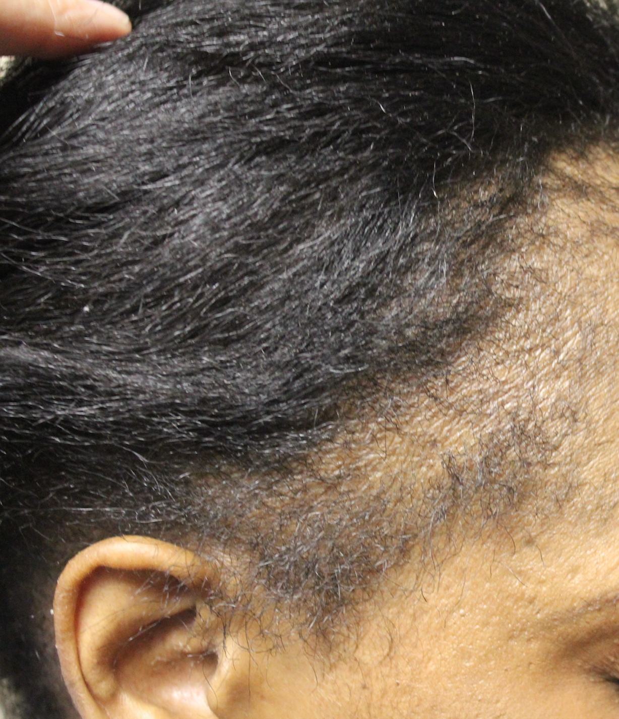
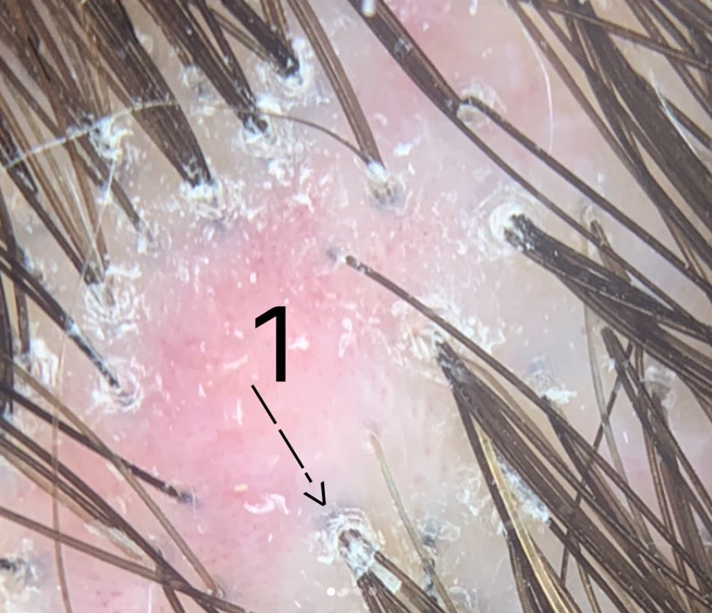
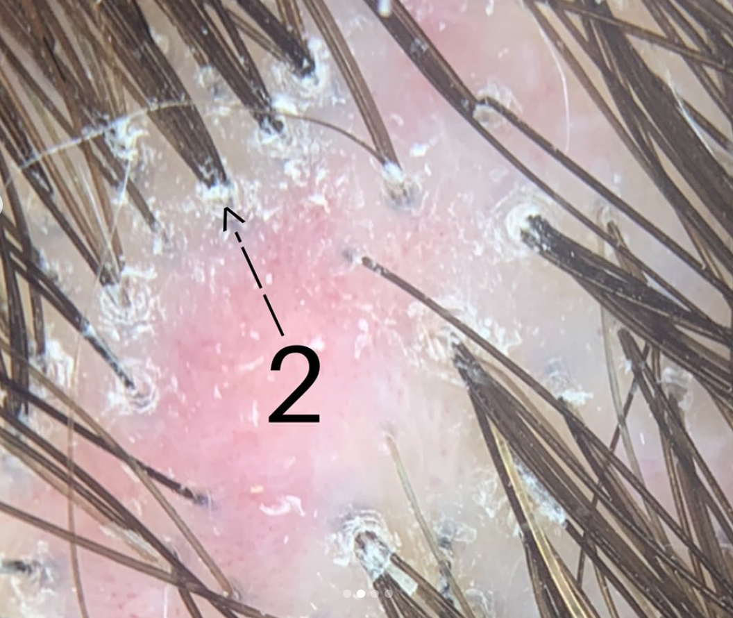
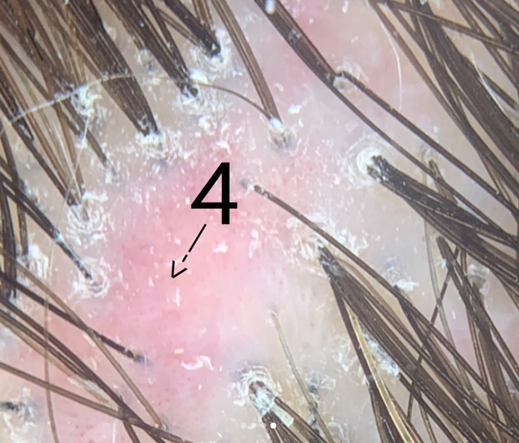
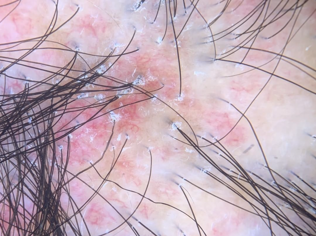
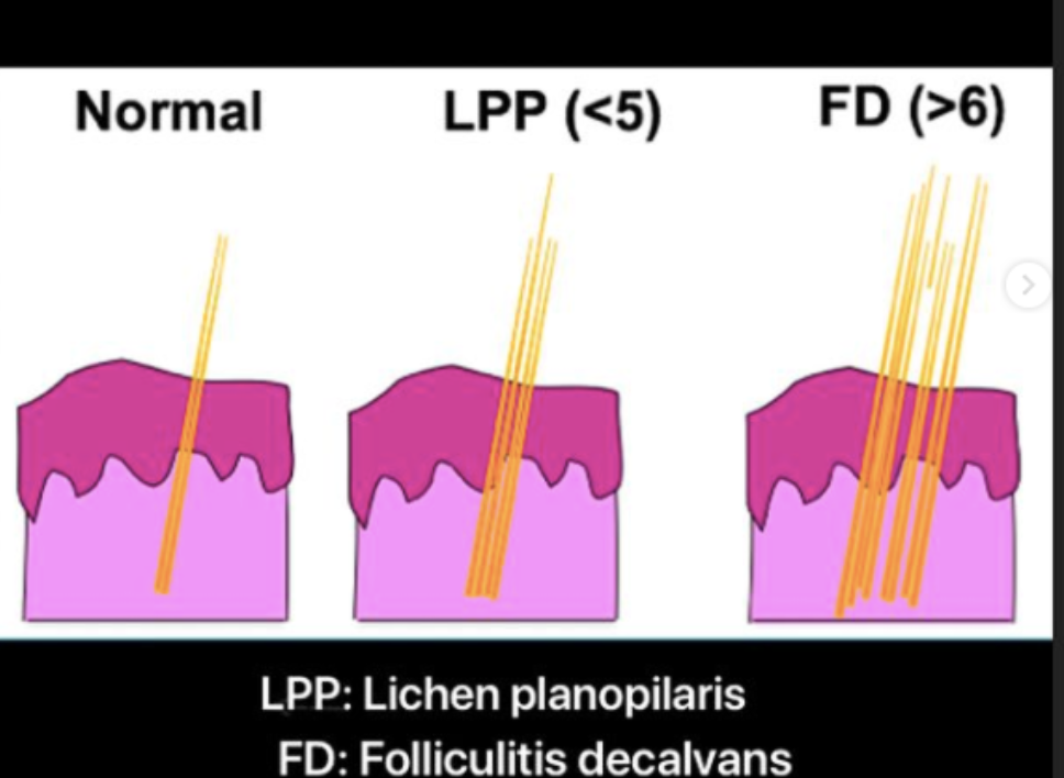
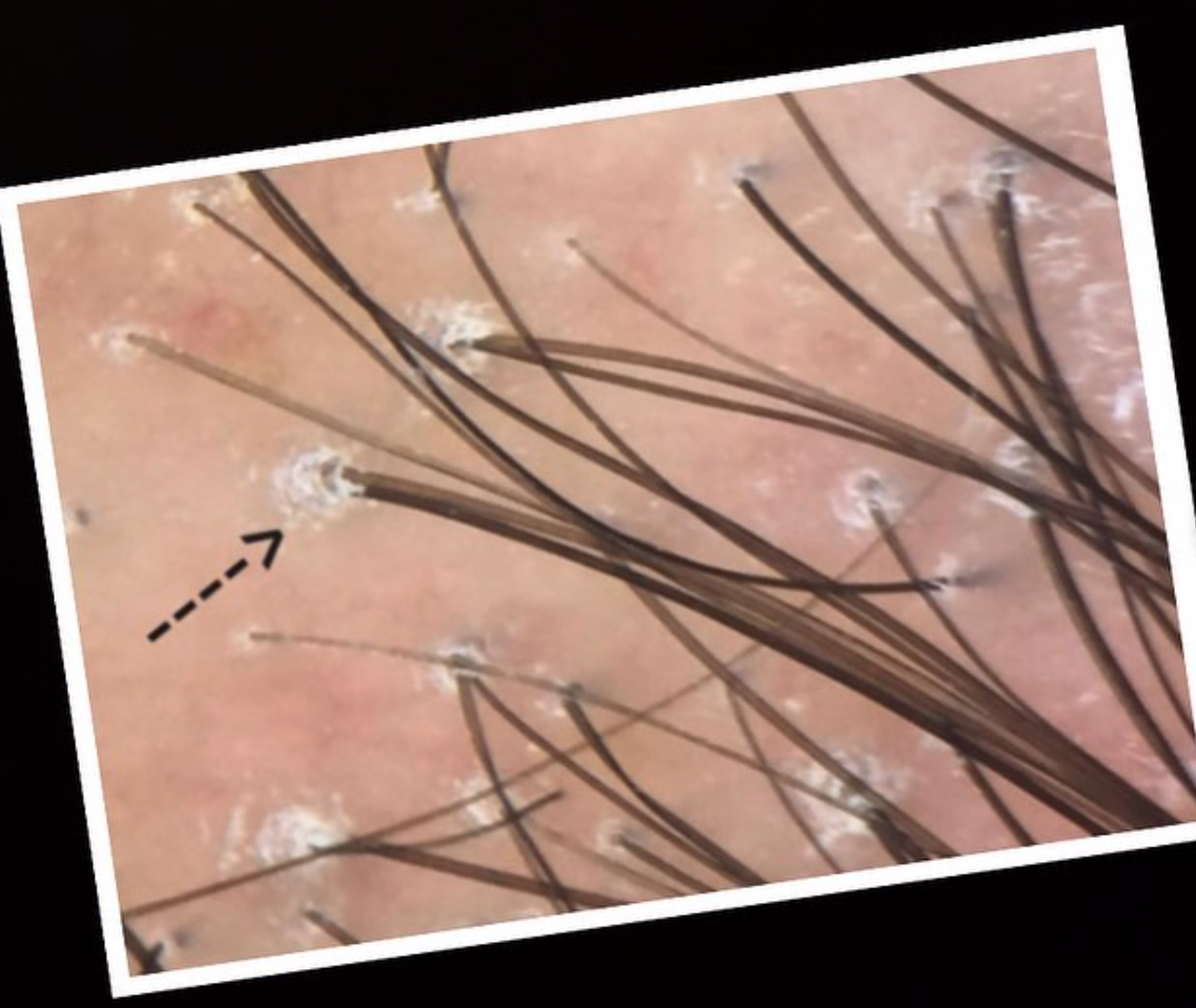
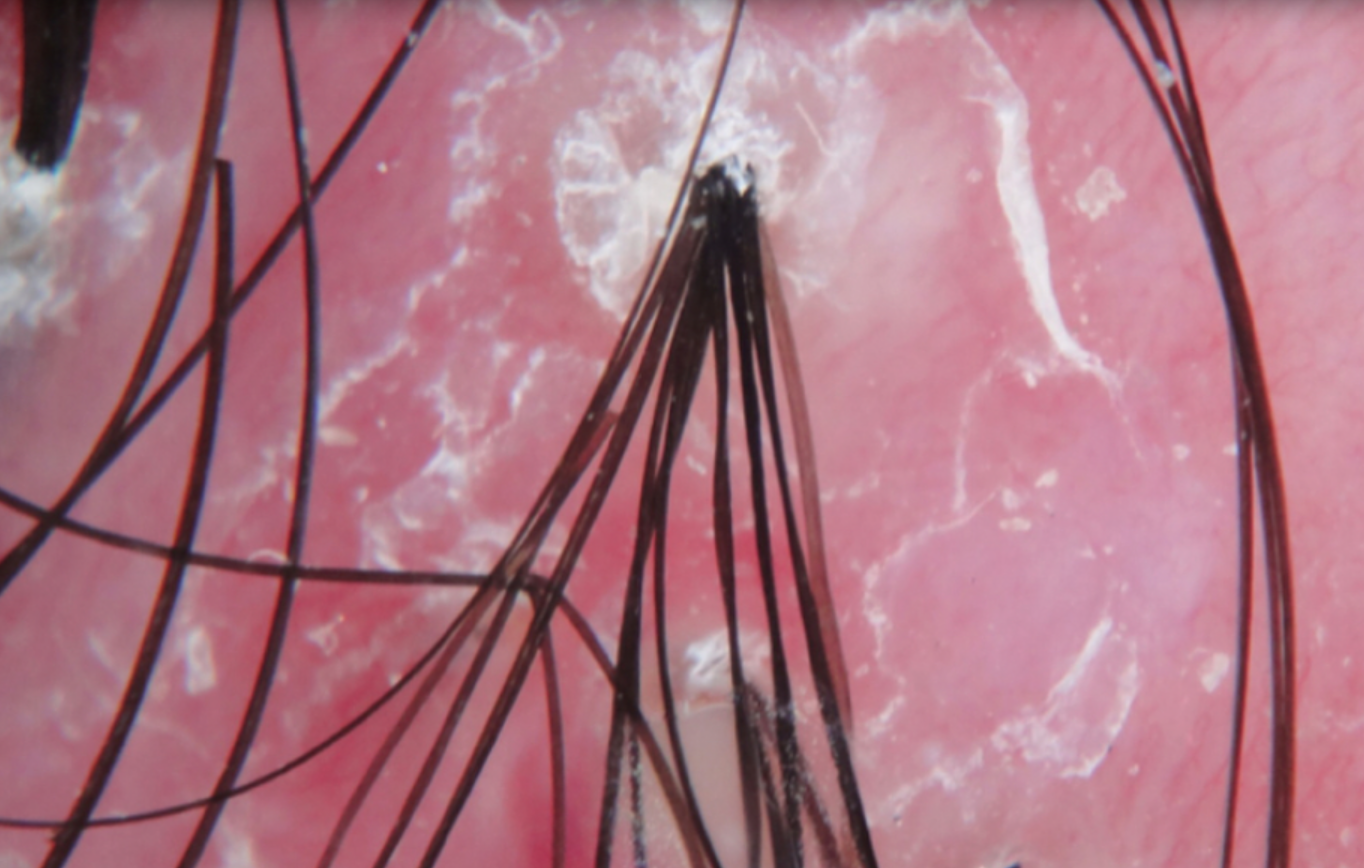
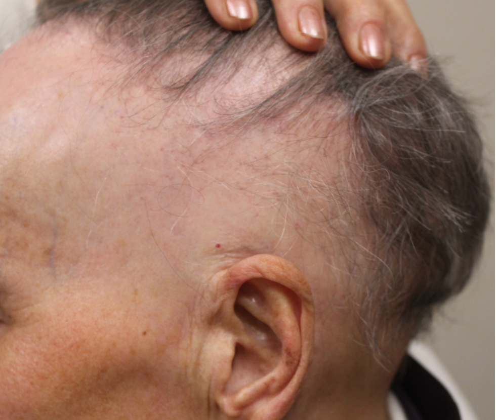
Folliculitis decalvans (FD) is a scarring alopecia that affects both men and women. FD is less common than some of the other  scarring alopecias like lichen  planopilaris and central centrifugal cicatricial  alopecia. The cause of FD is not completely know although a  role for bacteria has been postulated.   Bacteria such as Staphylococcus aureus frequently are found in the scalp of patients with folliculitis decalvans. Eradication  of bacteria with antibiotics, isotretinoin and other treatments frequently is associated with improvement of the disease.

A preliminary report proposes that Manuka honey may have benefits in the treatment of folliculitis  decalvans

A preliminary report proposes that Manuka honey may have benefits in the treatment of folliculitis decalvans


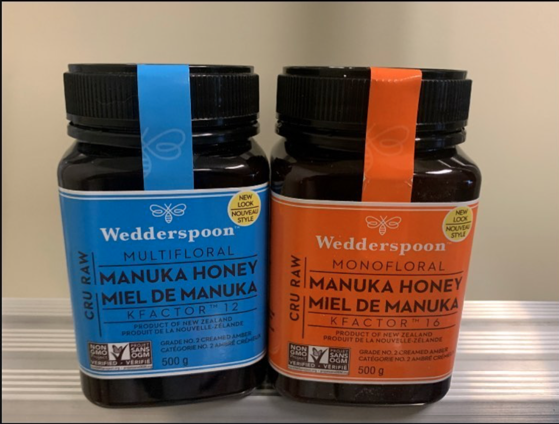
In 2019, a dermatology group in  Boston reported a patient with folliculitis decalvans whose disease improved with use of topical Manuka honey applied to the scalp. Manuka honeyis well known to wound care professionals. In fact, 17 clinical trial s involving  almost 2000 patients have suggested Manuka honey helps wounds heal.  Manuka honey has antibacterial properties – perhaps  due to its low pH, and other  plant  based and hydrogen peroxide based ingredients. A 1999 paper by Cooper and  colleagues showed that  Manuka honey killed  Staph aureus bacteria in wounds. 

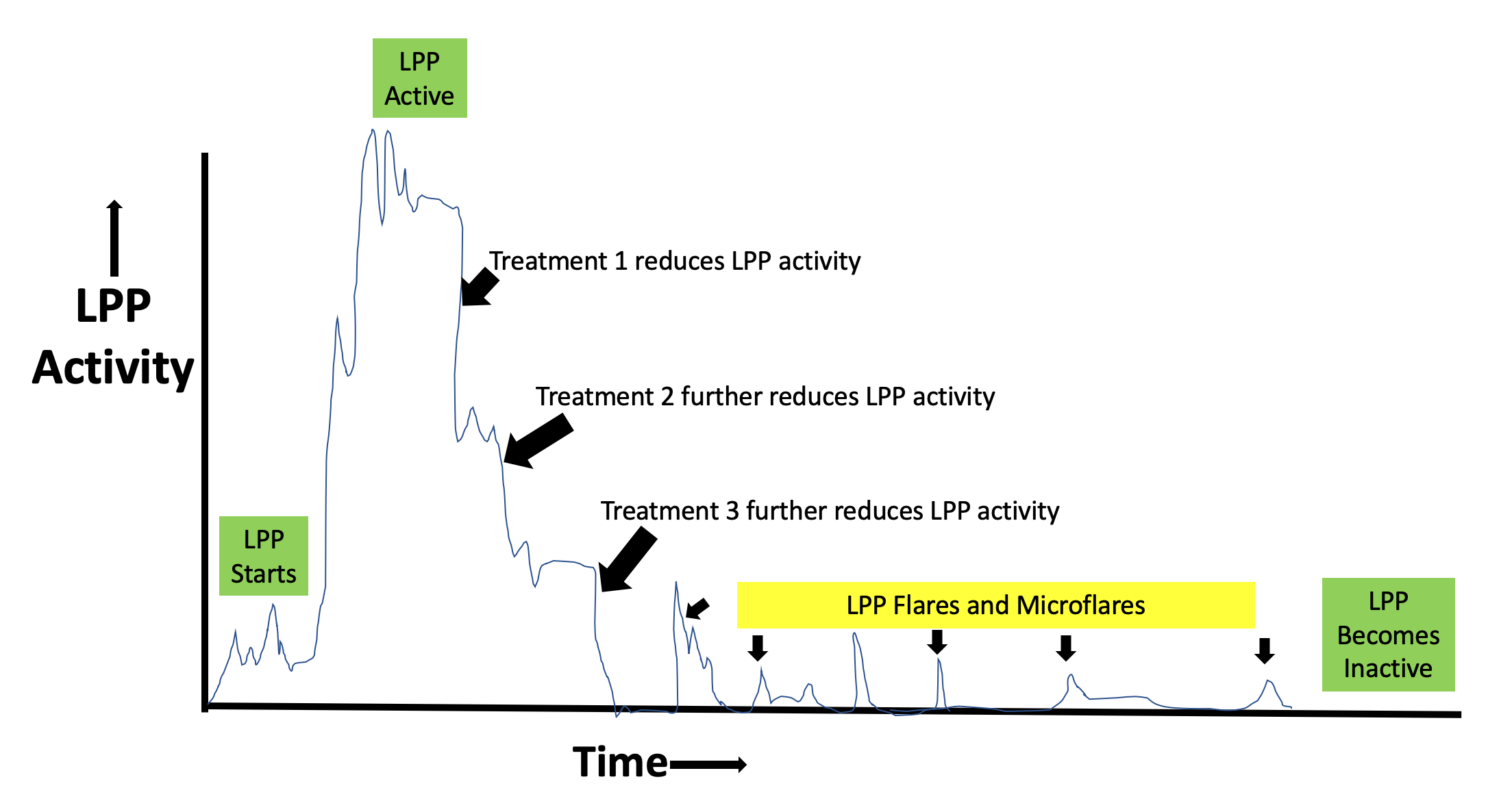
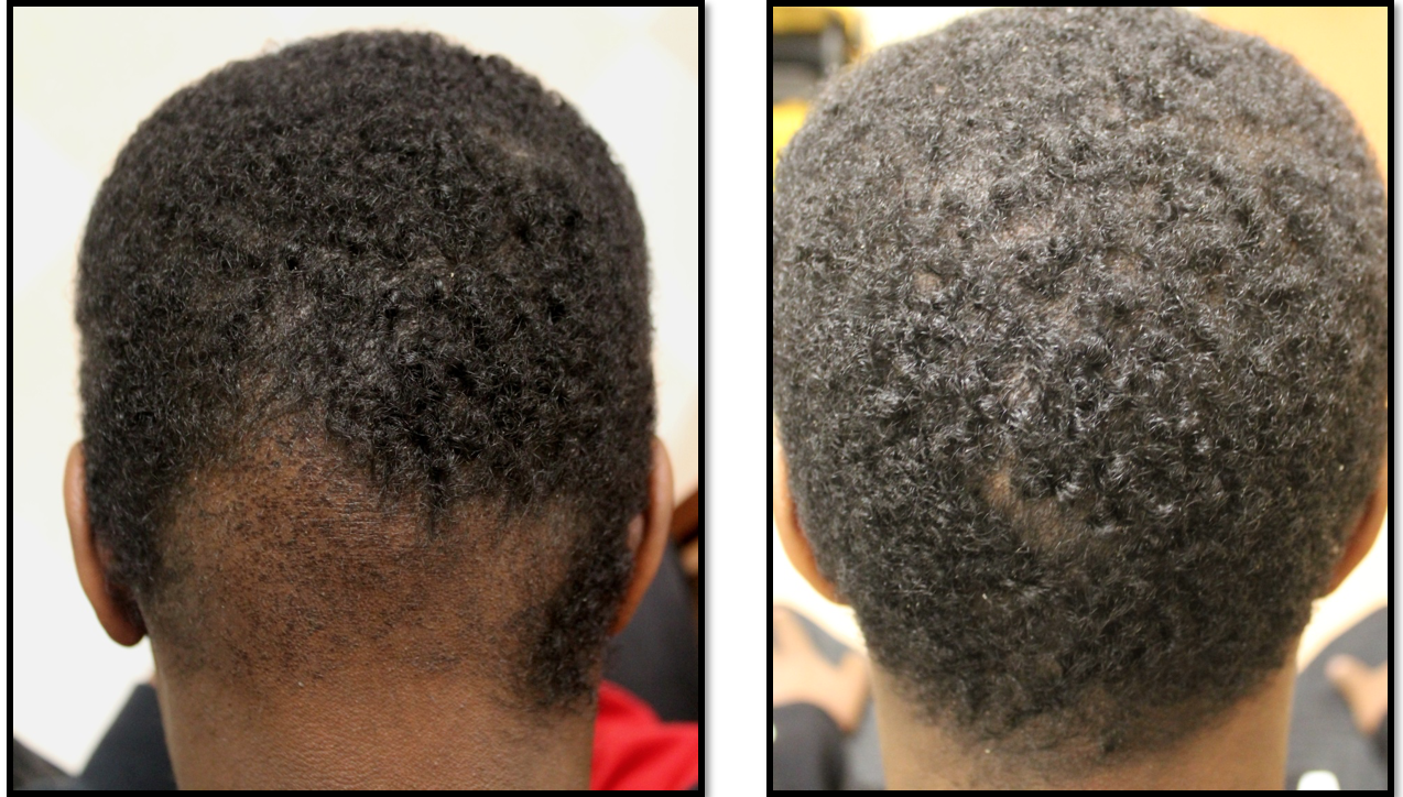
The patient in the 2019 paper was a 20 year old male who had used a considerable number of  treatment before he started applying manuka honey to his scalp. These treatments included steroid injections,  clobetasol lotion, prednisone, minocycline, doxycycline and isotretinoin. Some of these treatments were actually  quite helpful for the young man – however he had to  stop because of some side effects of these treatment.  

The  patient decided to start applying Manuka honey  to his scalp after about 1 month into a course of the oral antibiotic cephalexin. As he continued on both cephalexin and topical honey and found that after an additional 4 weeks of both treatments the scalp had improved considerably. 6 months later he stopped cephalexin. His disease eventually flared again and the man used honey alone to settle down his disease. 


Summary/Conclusion

This is an interesting paper. It doesn’t definitively prove Manuka honey helps FD but it hints that it might have a role. We also can’t rule out that the patients long term use of antibiotics and other treatments (like steroids) and isotretinoin have reduced disease activity that makes Manuka honey more likely to help. Nevertheless, this is an interesting paper that hopefully fuel more research in manuka honey.


Reference

Yeh et al. Resolution of folliculitis decalvans with medical honey. Dermatology Online Journal 2019;. 25(8); 


This article was written by Dr. Jeff Donovan, a Canadian and US board certified dermatologist specializing exclusively in hair loss.
Share This
No Comments

Cicatricial Marginal Alopecia: Your traction alopecia patients will thank you!

Not all ‘Traction Alopecia’ is Actually Traction Alopecia

Traction alopecia is a form of hair loss that occurs due to pulling of hair. Diagnosing traction alopecia sounds easy but surprisingly there are a great number of mimicking conditions that can fool the hair specialist.

Frontal traction alopecia refers to hair loss in the frontal hairline that is due to traction. Often the temples are affected but any part of the frontal hairline, temples and area around the ears can be affected. Often the hairs in the very frontal hairline are unaffected leading to the appearance of a so called “fringe” sign:

Classic ‘fringe’ sign in a patient with traction alopecia. The fringe refers to the fringe of hair in the frontal hairline.

Classic ‘fringe’ sign in a patient with traction alopecia. The fringe refers to the fringe of hair in the frontal hairline.

Cicatricial Marginal Alopecia (CMA)

There are times when patients who present with what seems to be traction alopecia tell us that they couldn’t possibly have traction alopecia. These are the patients who tell us that they have worn their hair fairly natural for years and that a diagnosis of traction alopecia just makes no sense to them. These are the patients that politely stare at us when we tell them to be careful how they style their hair and to be carefully to avoid heat or chemicals. When a hair specialist wants to make a diagnosis of traction alopecia but realizes the patient’s story just does not add up to give a convincing story of traction alopecia - the diagnosis of cicatricial marginal alopecia (CMA) must be considered.

The Differential Diagnosis of Frontal Hair Loss: What’s a specialist to consider anyways?

Of course, the diligent hair specialist considers many things in the differential of frontal traction alopecia like presentations including

1. Traction alopecia

2. Cicatricial Marginal Alopecia

3. Frontal fibrosing alopecia

4. Discoid lupus

5. Androgenetic alopecia

6. Telogen effluvium

7. Alopecia Areata

8. Trichotillomania

Cicatricial Marginal Alopecia: A Traction Alopecia Like Alopecia Without A Traction History

It was Dr Lynn Goldberg in Boston who put forth the notion of cicatricial marginal alopecia. She described 15 patients who presented with hair loss in a typical traction alopecia like pattern. Information pertaining to whether or not the patient relaxed or straightened the hair was available In 12 patients. 6 of the 12 patients gave a history of relaxing the hair or straightening the hair. For the other 6 other patients there was no such history. In other words, in 50 % of patients with frontal 'traction alopecia-like” hair loss a history of true traction styling practices were not present. These patients still had some degree of scarring on their biopsies indicating that this too could be a scarring type of hair loss. 

Treatment of CMA involves topical or oral minoxidil combined with topical and/or intralesional steroids. In some patients use of agents like oral doxycycline or topical tacrolimus can be helpful.

Summary and Key Lessons

As soon as we let open our mouths to pronounce the words traction alopecia, we must say in the same breath “or a traction alopecia like mimickers.” Could my patient have traction alopecia or a “traction alopecia like mimicker.”

Cicatricial Marginal Alopecia is one of these closely related mimickers. I like to refer to it as cicatrical marginal alopecia to honour my great colleague Dr Goldberg and so this is what I write in all my letters and consultation notes to other physicians. In my mind, I say the patient has a Traction Alopecia Like Alopecia Without a Traction History because it helps me remember the key elements of this presentation.

Reference

Goldberg L. Cicatricial Marginal Alopecia: Is It All Traction? Br J Dermatol 2009 Jan;160(1):62-8.


This article was written by Dr. Jeff Donovan, a Canadian and US board certified dermatologist specializing exclusively in hair loss.
Share This
No Comments

Scalp Trichoscopy: Completely Wonderful but Complete with Its Own Set of Limitations !

Can I just buy a USB trichoscope and figure out my own diagnosis ?

Trichoscopy is a wonderful diagnostic tool. Trichoscopy refers to the use of some sort of handheld device for viewing the scalp with higher magnification. These devices are widespread - they range in price from $ 35 for a pretty reasonable USB microscopy to $ 1,500 for a hand held device to $ 15, 000 for a video dermatoscope.

There are quite a few misconceptions that the pubic has about these devices.


1. Will trichoscopy tell me the diagnosis?

That answer is no. One can buy a USB device, plug it in and see beautiful pictures on the screen. But what does it mean? That requires an expert! It takes a few weeks to become reasonably good at trichoscopy and then a few years to become an expert. The USB trichoscope device does not give a print out that reads “you have androgenetic alopecia” or “you have telogen effluvium.”

Consider a useful analogy. If my air conditioner breaks down, I can certainly get out my tool box and open up the back of the air conditioner and see inside. But unless I known what I’m looking for, the process is not that useful and I will not know what’s wrong with the air conditioner (I can assure you based on my experience with doing this exact task).


2. If the trichoscope won't tell me the diagnosis, can’t I just email the doctor the pictures and he can tell me the diagnosis ?

I don’t like really ever answering two “no” answers in a row , but this answer is also no. We’re commonly asked this question. We have many people who ask us if they can just send in photos they have obtained with their own trichscope. These photos are not helpful UNLESS I have the entire story of the patient’s hair loss and have reviewed their blood tests and know absolutely everything about them. Then these trichoscopic images are a major bonus! It’s true that I can be pretty sure what’s going on by their photos - but not 100 % sure. Doesn't one want to be 100- % sure or at least as close to 100 % sure as possible?

The mistake people make is thinking trichoscopy is “everything.” They think to themselves that all I need to do is take pictures of my scalp of find some clinic to take trichoscopy pictures of my scalp and I’ll know what’s going on! That’s wrong, wrong wrong ….and that’s where I see people run into problems time and time again. Trichsocopy is wonderful but it’s only part of the puzzle. As an aside, some people also make the similar mistake of thinking that their blood test results are “everything.” They think to themselves that all I need to do is get to my doctor and get some blood tests and I’ll know what’s going on! That’s also not a correct approach. One needs the entire story and the chance to see the scalp in it’s entirely.

Although I’m sure I sound like a broken record, I’d like to remind the reader that the ideal way to diagnose hair loss is using what I termed the ”Diagnostic S.E.T.” I refer to these as the diagnostic “set” because theses 3 aspects all go together. These 3 items include:

1) the patient’s story

2) the findings uncovered during the process of the scalp examination (sometimes including trichoscopy, pull test, clinical exam, card test, etc)

3) the results of relevant blood tests. 

The first letter of each of the three words 1) story, 2) examination and 3) tests spell out the word “S.E.T.” - again a helpful reminder of how the information obtained from reviewing each of these 3 aspects helps solidify a proper diagnosis.

In summary, I can diagnose so many conditions with trichoscopy - but there are so many situations that I can not.

Let’s take a look at some situations where trichsocopy has it’s limitations.


EXAMPLE 1: TRICHOSCOPY IN THE NON SCARRING ALOPECIAS

Trichoscopy is completely wonderful. It helps me tremendously in the diagnosis of many non scarring alopecia. Most cases of androgenetic alopecia can be diagnosed with trichoscopy but not all! In fact, unless one is very experienced with trichscopy, the early cases of AGA are going to be very challenging to diagnose by trichscopy because there is just not enough miniaturization that has developed yet. So, if a patient buys a trichoscope and sees that their is not much miniaturization, can they conclude they don’t have AGA? No.

Most cases of acute alopecia areata can be diagnosed with trichoscopy. This is certainly one area where trichoscopy is very helpful. But in cases of advanced AA and some cases of alopecia areata incognito, all that might be seen is miniaturization of hairs. It can be difficult to render the diagnosis from trichscopy alone. So how do we diagnose it? Listen to the patient’s story!

Telogen effluvium (TE) refers to a type of hair shedding and is one of the more common diagnoses in women. Guess what? Telogen effluvium has NO definitive specific diagnostic trichoscopic signs ! Yikes! it’s true that the presence of many upright regrowing hairs can be a tip off from trichoscopy that the diagnosis of TE might be present - but it’s not specific. If a person thinks they are going to diagnose their TE by buying a trichoscope, they are wrong.

trichoscopy in TE- limitations


EXAMPLE 2: TRICHOSCOPY IN THE SCARRING ALOPECIAS

Trichoscopy is completely wonderful. It helps me tremendously in the diagnosis of many scarring alopecia. In fact, the use of trichoscopy has massively reduced my need to perform scalp biopsies. That said, one needs to be aware that some cases of early lichen planopilaris can’t be confidently diagnosed with trichoscopy - the scalp looks just like seborrheic dermatitis! Some cases of early folliculitis decalvans look just like regular ordinary folliculitis !

So does trichosopy help in all these subtle and early forms of these diseases? - no ! It gets me thinking but usually a biopsy is needed to confirm these challenging diagnoses.

Let it be heard though - a good majority of scarring alopecia cases can be diagnosed with trichosopy. Just not all!

As for central centrifugal cicatricial alopecia (CCCA), the best way to diagnose this condition is simply to look at the scalp! Trichoscopy can help but there are not a great number of classic trichscopic signs for CCCA.

trichoscopy scarring alopecia


FINAL SUMMARY

Many patients want to get blood tests because they think that the blood tests will provide the entire answer the diagnosis. Many patients want to buy a trichoscope (USB dermatoscope) because they feel the trichoscope will provide the answers.

We must always remember that the confident diagnosis of hair loss from from use of the diagnostic SET - all comments from the patient’s story, scalp examination,, trichoscopic examination and blood tests go into figuring out the exact cause.


This article was written by Dr. Jeff Donovan, a Canadian and US board certified dermatologist specializing exclusively in hair loss.
Share This
No Comments

Trichoscopy of Folliculitis Decalvans

Key Trichoscopic Signs of Folliculitis Decalvans

Folliculitis decalvans (FD) is a scarring alopecia which causes permanent hair loss. Patients develop red, itchy scalps that often contains pimples. Bacteria such as Staphylococcus aureus can sometimes be isolated when swabs are taken from these pimples.

A number of “trichoscopic” or “dermatoscopic” signs are suggestive of folliculitis decalvans including some I have shown here: (1) perifollicular “tubular” scaling, (2) compound follicles containing 6 or more hairs, (3) linear fibrotic bands and the (4) red “strawberry ice cream” color.

(1) Perifollicular “tubular” scaling

Trichoscopy (Scalp Dermoscopy) of Folliculitis Decalvans, Image 1. tubular scaling. The scale rides up higher on the hair shaft in folliculitis decalvans than in lichen planopilaris.

Trichoscopy (Scalp Dermoscopy) of Folliculitis Decalvans, Image 1. tubular scaling. The scale rides up higher on the hair shaft in folliculitis decalvans than in lichen planopilaris.

(2) Compound Follicles (Containing 6 or more Hairs)

Trichoscopy (Scalp Dermoscopy) of Folliculitis Decalvans, Image 2. “Compound follicles” are follicles containing more than 6 hairs emerging from a single pore.

Trichoscopy (Scalp Dermoscopy) of Folliculitis Decalvans, Image 2. “Compound follicles” are follicles containing more than 6 hairs emerging from a single pore.

(3) Linear fibrotic bands

Trichoscopy (Scalp Dermoscopy) of Folliculitis Decalvans, Image 3. Linear Fibrotic bands indicate a pattern of scarring associated with the typical starburst scaling.

Trichoscopy (Scalp Dermoscopy) of Folliculitis Decalvans, Image 3. Linear Fibrotic bands indicate a pattern of scarring associated with the typical starburst scaling.

(4) Red “Strawberry ice cream” Color.

Trichoscopy (Scalp Dermoscopy) of Folliculitis Decalvans, Image 4. Strawberry Red Color.

Trichoscopy (Scalp Dermoscopy) of Folliculitis Decalvans, Image 4. Strawberry Red Color.


Folliculitis decalvans (FD) vs Lichen planopilaris (LPP).

Folliculitis decalvans can resemble lichen planopilaris at first glance. However, it does have many differences. Compared to LPP, FD is more likely to have pustules, is more likely to bleed, is more likely to showing compound follicles or “tufting” and is more likely to have tubular scaling the climbs up the follicles (as in this image) and more likely to have these linear fibrotic bands too. Treatment for FD has been discussed in other posts but includes antibiotics, isotretinoin as well as other treatments. 


This article was written by Dr. Jeff Donovan, a Canadian and US board certified dermatologist specializing exclusively in hair loss.
Share This
No Comments

Lichen planopilaris: Classic trichoscopic findings

Classic trichoscopic findings of LPP

Classic trichoscopy of active lichen planopilaris, an immune mediated scarring alopecia is shown below.

Classic trichoscopic image from a patient with active lichen planopilaris (LPP)

Classic trichoscopic image from a patient with active lichen planopilaris (LPP)

There is redness and scale around hairs (called perifollicular erythema and perifollicular scale). Some hairs are twisted (called pili torti). The areas of scalp devoid of hairs are no longer red as the immune system has destroyed hairs in that area and has since left the area. Treatments discussed in other posts as in the following link.

Treatments for LPP: What is available?


This article was written by Dr. Jeff Donovan, a Canadian and US board certified dermatologist specializing exclusively in hair loss.
Share This
No Comments

The Course of Treating Lichen Planopilaris: What do we hope for?

A Closer Look at the Typical Course of Treating Lichen Planopilaris

Lichen planopilaris is a scarring alopecia. Treatments are largely focused on reducing inflammation in hopes that by doing so the disease can be stopped. 


In the present day, there are many aspects of LPP that are difficult to predict. We know to some degree which treatments are better than others. We know the proportion of patients that are expected to benefit from a certain type of treatment and the proportion of patients who are not expected to benefit. However, we do not know if a given patient will require several trials of medications before finding one that ‘works’ to suppress the disease and whether a given patient will need 1, 2 3 or more treatments to suppress the disease completely (and therefore stop it). In addition, we do not know how long the patient will need to use their treatments before the disease completely stops. It ranges from a year or two to many decades.

When all goes well, LPP disease activity eventually settles down with use of one or more medications. Disease flares and micro flares can still happen even when patents are on the right treatment plan. Some patients, but not all, finally enter a sta…

When all goes well, LPP disease activity eventually settles down with use of one or more medications. Disease flares and micro flares can still happen even when patents are on the right treatment plan. Some patients, but not all, finally enter a state where their disease is completely quiet. When the disease stays quiet and mediations can be stopped, we call the condition “burnt out.”


Flares and MIcroflares in LPP

Even when a patient’s disease achieves a fairly inactive state, flares and microflares may still happen from time to time.  These refer to sudden increases in the activity of the disease. Patients who are experiencing flares may become more itchy, or notice more daily hair shedding. Stress, heat, can humidity can cause such microflares and sometimes these sorts of flares occur for no apparent reason at all.Some patients, but not all, finally enter a state where their disease is completely quiet. When the disease stays quiet and mediations can be stopped, we call the condition “burnt out.” 

 Although no patient is really ‘typical’ when it comes to LPP, a standard treatment protocol is summarized in this graph above.


This article was written by Dr. Jeff Donovan, a Canadian and US board certified dermatologist specializing exclusively in hair loss.
Share This
No Comments

Challenge Case of Compound Follicles - Final Diagnosis Lichen planopilaris

The Case of Compound Follicles

“Compound follicles” are follicles with 6 or more hairs coming out of a single pore. Occasionally, follicles with 4,5 and rarely 6 hairs coming out of a single pore can be found as an isolated finding- especially towards the more posterior regions of the scalp. But large groupings of hairs like this are rare with advancing age and in fact - the presence of many areas containing compound follicles should prompt the clinician to consider that the patient might have a scarring alopecia. Compounding occurs when 2 or more adjacent follicles “fuse” together. They do not occur because a hair follicle suddenly makes more hairs. Compound follicles are more common in neutrophilic scarring alopecias like folliculitis decalvans (FD) than in lymphocytic scarring alopecias like lichen planopilaris (LPP). Nevertheless, occasional follicles with 5 and 6 hairs can be found in LPP…. as we’ll see in the case below.

I’ve included below a schematic diagram showing the typical findings in lichen planopilaris (LPP) and folliculitis decalvans (FD). Lichen planopilaris typically has no compounding although rarely it’s true that we can see it ….as we’ll seen in the case below. Compounding is more common in folliculitis decalvans.

compound follicles


Generally speaking, the tendency in LPP is for hairs to be destroyed and follicles to contain fewer and fewer hairs over time rather than to contain more follicles in them.

Case

case 1


The case was a patient who presented with redness in the scalp and a loss of hair density. Trichosocpy of the scalp is shown in the photo above. The arrow points to a follicle with 6 hairs coming out of a single pore. There were not many other features in this photos that suggested LPP such as perifollicular scale or perifollicular erythema or pili torti or scarring.

Below is a more typical photo of lichen planoplaris. As you’ll likely agree, the above photo doesn’t quite look like the only below. The typical photo of LPP has scale around hairs. Furthermore, most of the hairs either come out of the pores in groups of 2 or just one hair or no hair at all !

Typical trichoscopic image of lichen planopilaris. Perifollicular scale (white scare around hairs) is evident.

Typical trichoscopic image of lichen planopilaris. Perifollicular scale (white scare around hairs) is evident.


Occasional hairs in our patient’s case had a thicker scale than expected in LPP which prompted me to also consider whether this could be “starburst” scaling of FD. A “hint” of crusting is present but there are no pustules. Redness is interfollicular. A typical trichoscopic image of FD is shown below:

Typical trichoscopic image of folliculitis decalvans. Starburst scale is seen around hairs and compound hairs (hairs with more than 6 hairs coming out of a single follicular opening) are clearly evident.

Typical trichoscopic image of folliculitis decalvans. Starburst scale is seen around hairs and compound hairs (hairs with more than 6 hairs coming out of a single follicular opening) are clearly evident.


A biopsy was be done to evaluate for the possibility of scarring alopecia. The biopsy returned showing lichen planopilaris with no features of folliculitis decalvans. The biopsy also showed that there was a perifollicular inflammatory infiltrate of lymphocytes together with lichenoid change (death of keratinocytes) in the outer root sheath. Perifollicular fibrosis was seen along with loss of sebaceous glands.

FINAL DIAGNOSIS: LICHEN PLANOPILARIS. 

Comment on Case

This was a nice example of a case that was atypical. Not all patients with lichen planopilaris have a typical presentation. Most however have scalp symptoms (like itching or burning or tenderness). Most have redness of some sort in the scalp. Most of scale around hairs. This patient had a bit of subtle redness and not really that much in the way of symptoms. The patient had some unusual compounding by trichoscopy which was the tip off that something might not be right. The biopsy confirmed the diagnosis of lichen planopilaris. Compounding is not a typical feature of LPP but certainly can be seen from time to time. It’s usually not a feature seen in all regions of the scalp and usually the compounding is limited to less than 7 hairs. Compound hairs containing 10, 15 our 20 hairs are almost never seen in lichen planopilaris (LPP) but can be seen very commonly in folliculitis decalvans (FD).

The patient was started on topical steroids and steroid injections together with hydroxychloroquine. The patient will be seen back in 3 months to review response to treatment. Blood tests will be needed monthly for three straight months for CBC, AST, ALT while starting hydroxychloroquine. An eye examination will be needed within 6 months. Clinical photos and trichoscopic photos were taken at the first visit and wil be compared to photos taken a the 3 months follow up. The hope is that redness will be reduced and that that patient’s perception of increased shedding will be reduced. I will monitor over time if more hair loss occurs. Regrowth may or may not occur in scarring Alopecias and this is not a main goal. The goal of treatment is to stop it from getting worse.


This article was written by Dr. Jeff Donovan, a Canadian and US board certified dermatologist specializing exclusively in hair loss.
Share This
No Comments

Steroid Injections for Hair Loss - A Look at Triamcinolone Acetonide

Steroid Injections with Triamcinolone Acetonide

Steroid injections are extremely helpful for many hair loss conditions - particularly some forms of localized alopecia areata (AA) some patients with lichen planopilaris (LPP), frontal fibrosing alopecia (FFA), central centrifugal cicatricial alopecia (CCCA) and folliculitis decalvans. I sometimes even use in the early stages of traction alopecia when I feel the condition is in its earliest stages.

Triacminolone acetonide is a common medication used for steroid injections for inflammatory related causes of hair loss. Kenalog is a popular brand name and available in 10 mg and 40 mg bottles. The final dose the physician uses is typically 2.5 to …

Triacminolone acetonide is a common medication used for steroid injections for inflammatory related causes of hair loss. Kenalog is a popular brand name and available in 10 mg and 40 mg bottles. The final dose the physician uses is typically 2.5 to 5 mg per mL.


There are two common doses of triamcinolone acetonide that one orders from the manufacturer - 10 mg per mL and 40 mg per mL. Either is fine to order provided one keeps in mind that when using the 40 mg per mL dose one is going to need to use 4 times less than if using the 10 mg per mL dose. Every few months I get calls from physicians who call me in a panic because they have prepared their injections using a 40 mg per mL bottle but they thought it was a 10 mg per mL bottle. (the correct way to deal with this is to 1) admit one’s error to the patient, and then 2) flood the scalp generously with saline injections to dilute out the steroid and see the patient back in 4 weeks and 8 weeks to see if any atrophy developed). There are many brands of triamcinolone acetonide one can order. Kenalog is one brand (shown here) but there are others. I have used many over the years and find some do get clogged up when using tiny 30 gauge needles. I don’t find this happens with Kenalog.

As reviewed in other posts, I believe in starting steroid injections at 2.5 mg per mL and only going to 5 mg per mL if needed. The low dose can be helpful and allows more injections to be performed as the maximum dose is 4 mL if one uses a 5 mg/mL concentration or 8 mL if one uses a 2.5 mg per mL concentration.


This article was written by Dr. Jeff Donovan, a Canadian and US board certified dermatologist specializing exclusively in hair loss.
Share This
No Comments

Cetirizine (Zyrtec) in Lichen Planopilaris: Best Viewed as an Adjunct not Main Treatment

Cetirizine (Zyrtec) for Treating Lichen Planopilaris: Where does it fit in?

Antihistamines are increasingly being studied for the treatment for various types of hair loss. For the autoimmune disease known as alopecia areata for example, use of antihistamines like fexofenadine (Allegra) as well as others (i.e. ebastine) may have some treatment related benefits. In scarring alopecia. the use of antihistamines has only received a limited amount of study. Today, we will discuss the use of the antihistamine cetirizine for treating lichen planopilaris.

Lichen planopilaris is an immune medicated disease. There are a number of proposed mechanisms that lead ultimately to the disease. Cetirizine is an antihistamine medication and widely used for various types of allergy related symptoms. However, the medication may have a number of general and wide reaching effects on the immune system.


How does cetirizine work and how does it affect the immune response ?

Cetirizine is an H1 receptor antagonist. The drug minimally crosses the so called ‘blood brain barrier’ and so limited amounts actually get into the brain. This results in less sedation with cetirizine compared to any other traditional antihistamines. The 5 mg and 10 mg doses are unlikely to give sedation for most people. However, the 20 mg and 30 mg doses are much more likely to give sedation. There has been concern in recent years among long term chronic use of high doses of antihistamines on cognitive decline in patients so this needs to be taken into account when discussing high dose cetirizine as chronic therapy with patients with any medical condition.


Cetirizine has a number of potential effects to modify the immune response. These include

1. Inhibit DNA binding activity of NF-kappa B,

2. Inhibit the expression of adhesion molecules on immunocytes and endothelial cells

3. Inhibit the production of IL-8 and LTB4, two potent chemoattractants, by immune cells.

4. Induce the release of PGE2, a suppressor of antigen presentation and MHC class II expression, from monocyte/macrophages

5. Reduces the number of tryptase positive mast cells in inflammation sites.


The 2010 d’Ovidio Lichen Planopilaris Study

In 2010, d’Ovidio and colleagues studied the use of cetirizine at high doses. Rather than using 5 mg to 10 mg daily that is commonly use over the counter, the authors studied the benefits of 30 mg/daily. Twenty-one patients with lichen planopilaris (LPP) were treated with cetirizine as well as their topical steroids. in 18 or 21 patients (85.7 %) there was a reduction in redness, scaling and a reduction in extractable anagen hairs by the pull test. The authors reported that one patient developed cardiac arrhythmia after 3 months of successful treatment and dropped out of the study.

Cetirizine is an antihistamine and functions as an H1 receptor antagonist. In 2010, d’Ovidio showed that cetirizine at high doses (30 mg) could benefit some patients with lichen planopilaris. Over the counter antihistamine dosing like shown in the p…

Cetirizine is an antihistamine and functions as an H1 receptor antagonist. In 2010, d’Ovidio showed that cetirizine at high doses (30 mg) could benefit some patients with lichen planopilaris. Over the counter antihistamine dosing like shown in the phone is 10 mg.


What are the side effects of cetirizine?

Side effects of cetirizine and other information can be found in our Handout.

Cetirizine Handout for LPP

Rare side effects including heart failure, angioedema and tachycardia. These side effects are rare at low doses such as the 5 mg and 10 mg (over the counter doses). Side effects increase as one increased the dose. The 30 mg dose used in the d’OIividio study would be expected to have a greater degree of side effects than the lower doses.


Conclusion

Cetirizine may have some benefit in treating lichen planopilaris. I sometimes prescribe cetirizine as an adjective treatment in patients with persistent itching and burning who are not fully responding to mainstay topical, intralesional and oral treatments. Generally I use 5 mg or 10 mg and only rarely do I prescribe 15-20 mg. I do not typically prescribe 30 mg doses as I find side effects increase greatly. One must respect the drug interactions and contraindications for the drug (as outlined in the handout). In many ways, I view cetirizine as a helpful add on - much the same way as I view the use of low level laser therapy in this disease. I do not think in the present day that cetirizine should find itself at the top of the therapeutic ladder but certainly has a place.


Reference

d’Ovidio R et al Therapeutic hotline. Effectiveness of the association of cetirizine and topical steroids in lichen planus pilaris--an open-label clinical trial. Dermatol Ther. 2010 Sep-Oct;23(5):547-52.


Namazi MR et al. Cetirizine and allopurinol as novel weapons against cellular autoimmune disorders.Int Immunopharmacol. 2004 Mar;4(3):349-53.


This article was written by Dr. Jeff Donovan, a Canadian and US board certified dermatologist specializing exclusively in hair loss.
Share This
No Comments

Is Immunotherapy (DPCP, SADBE) Effective for Lichen Planopilaris or Frontal Fibrosing Alopecia ?

Immunotherapy (DPCP, SADBE) Does NOT Help Scarring Alopecia

Diphencyprone (DPCP) and Squaric Acid Dibutyl Ester (SADBE) are long standing treatments for the autoimmune hair loss condition alopecia areata. The cause itching and burning in the scalp and essentially trigger an allergic contact dermatitis. The inflammation that these chemicals create can trigger hair growth in some patients with alopecia areata. It’s quite remarkable.

FIGURE 1. Diphenycyprone (DPCP) is a liquid that is applied to the scalp to treat alopecia areata. It causes an allergic reaction but can stimulate hair growth in some users.

FIGURE 1. Diphenycyprone (DPCP) is a liquid that is applied to the scalp to treat alopecia areata. It causes an allergic reaction but can stimulate hair growth in some users.

FIGURE 2: Hair Regrowth in a patient with ‘ophiasis” type of alopecia areata who was treated with diphenyprone.

FIGURE 2: Hair Regrowth in a patient with ‘ophiasis” type of alopecia areata who was treated with diphenyprone.


Does DPCP and Squaric Acid Help Lichen Planopilaris or Frontal Fibrosing Alopecia (FFA)?

DPCP and Squaric acid are not effective in these scarring alopecias. It’s not that large studies have been done - it’s just that I’ve seen patients many patients over the years with scarring alopecias who have come to see my after having DPCP. These patients were all mistakenly diagnosed as having alopecia areata when really they had frontal fibrosing alopecia or lichen planopilaris. Hair regrowth did not occur and many experiencing a significant worsening.

FIGURE 3: Patient with frontal fibrosing alopecia who was first thought to have the ophiasis form of alopecia areata and was treated with DCPC for many months. The patient did not experience regrowth.

FIGURE 3: Patient with frontal fibrosing alopecia who was first thought to have the ophiasis form of alopecia areata and was treated with DCPC for many months. The patient did not experience regrowth.

There is absolutely no reason to believe that DPCP or squaric acid are effective in LPP or FFA. The pathogenesis of these two conditions is very different than alopecia areata. While it’s true that some treatments overlap - many do not. The following table summarizes some of these important differences. DPCP is effective for some patients with alopecia areata but is not effective in LPP. Doxycycline is effective for some patients withLPP but not helpful in treating alopecia areata.

Table 1: Treatments in Alopecia Areata vs Lichen Planopilaris

AA vs LPP

This article was written by Dr. Jeff Donovan, a Canadian and US board certified dermatologist specializing exclusively in hair loss.
Share This
No Comments

Patient-Doctor Conference Focused on Scarring Alopecia

CARF’s Patient-Doctor Conference All Set For Nashville, TN

I’m looking forward to another great meeting of the Cicatricial Alopecia Research Foundation (CARF). Every 2 years, CARF hosts a wonderful meeting that brings together patients and physicians. I’ve attended for several years now and always enjoy it. I’ll be speaking again at this year’s meeting and look forward to seeing everyone there. The two and a half day meeting is packed with lots of great information, support, and good fun.

Information on the meeting can be found on the CARF website and is also highlighted here:

CARF 2020 Patient Doctor Conference

CARF Prg

The conference brochure can be downloaded here:

CARF 2020 in Nashville, TN: April 24-26, 2020

carf 2020


This article was written by Dr. Jeff Donovan, a Canadian and US board certified dermatologist specializing exclusively in hair loss.
Share This
No Comments

The Use Metformin in Lupus: How does this apply to our Discoid LE (DLE) Patients?

Metformin for Lupus: Will it help discoid lupus?

Metformin is a well known diabetes drug. Recent evidence has suggested that metformin may have a positive impact on the treatment of some autoimmune diseases.

Metformin is well understood to reduce glucose production by the liver and to reduce absorption of glucose in the gastrointestinal tract and to increase insulin sensitivity. However, metformin may also reduce production of reactive oxygen species (ROS) which help create inflammation in lupus. The possible benefits of metformin in lupus was demonstrated in 2015 where Wang and colleagues in the journal Arthritis and Rheumatolgy showed that metformin reduce the risk of disease flares by 51 % compared to conventional treatment.

In a 2018 poster by McLeod and colleagues presented at the 2018 meeting of the American College of Rheumatology, authors showed that metformin helps patients with lupus improve control of their disease. The researchers studied 15 patients with lupus using metformin and compared to 1331 patients not using metformin. The authors found there ws a difference in disease activity in patients using metformin.

Metformin for Discoid Lupus: Will it help?

These studies are interesting as they suggest that metformin has the potential to help patients with lupus. What we don’t know yet is whether metformin will help the various types of ‘cutaneous’ lupus including discoid lupus.

Metformin may have an impact not only on autoimmune diseases (including effects on monocytes, macrophages and neutrophils), but improve gut microbiota and have an antifibrotic effect as well. These effects together make them ideal to consider in the study of scarring alopecia. We already know that drugs as pioglitazone may be helpful in lichen planopilaris.

REFERENCES

Wang et al. Neutrophil Extracellular Trap Mitochondrial DNA and Its Autoantibody in Systemic Lupus Erythematosus and a Proof-of-Concept Trial of Metformin.Arthritis Rheumatol. 2015 Dec;67(12):3190-200. doi: 10.1002/art.39296.

McLeod C, Olayemi G, Bhatia N, Migliore F, Quinet R. The Impact of Metformin on Disease Activity in Systemic Lupus Erythematosus [abstract]. Arthritis Rheumatol. 2018; 70 (suppl 10). https://acrabstracts.org/abstract/the-impact-of-metformin-on-disease-activity-in-systemic-lupus-erythematosus/. ABSTRACT NUMBER: 2645


This article was written by Dr. Jeff Donovan, a Canadian and US board certified dermatologist specializing exclusively in hair loss.
Share This
No Comments

Frontal Fibrosing Alopecia: The Early Stages

When FFA First Begins 

FFA-early

Frontal fibrosing alopecia (FFA) is a scarring alopecia that affects women to a greater extent than men. The cause remains unknown although hormonal and immune-based mechanisms are clearly relevant.

The disease causes loss of hairs in the frontal hairline, sides and back of scalp, eyelashes, eyebrows and body hair. What is interesting about FFA is that the very earliest stages are associated with destruction of the tiny “vellus” hairs. This destruction leaves behind the thicker terminal hairs. 


In the earliest stages of FFA, the hair loss can be completely unnoticeable. There are frequently no symptoms and there is simply a subtle thinning in the area rather than complete loss. 

This photo of a patient with FFA shows a relatively normal looking scalp that is easily mistaken for androgenetic alopecia. (In fact this photo could easily be a picture of androgenetic alopecia were it not for the loss of all vellus hairs in this area over a 3 month period). In androgenetic alopecia, there is a gradual (slow!) conversion of thick hairs to thin hairs (a process called miniaturization). In FFA, we often do not see the miniaturized and vellus hairs as they are preferentially destroyed by the immune system. We see mainly single terminal hairs in FFA. Over time (without treatment) there may be some redness that develops in this area and even some scaling. About 40 % of women with FFA have androgenetic alopecia as well, so the two conditions frequently co-exist.

It is often not a decision “is this FFA or AGA ...but rather is it FFA, AGA or both.” The goal of treatment however is to stop that from occurring and the patient was started on topical fluocinonide gel, pimecrolimus cream, steroid injections and oral finasteride.


This article was written by Dr. Jeff Donovan, a Canadian and US board certified dermatologist specializing exclusively in hair loss.
Share This
No Comments

Characteristics of FFA in Men

 Characteristics of FFA in Men

Frontal fibrosing alopecia is a type of scarring alopecia that causes hair loss along the frontal hairline and sideburns but can also affect the back of the scalp, eyebrows, eyelashes and body hair.  For every 100 patients I see with a diagnosis of FFA, 99 patients are women and 1 patent is   male.

Tolkachjov and colleagues performed a study of 7 male patients with frontal fibrosing alopecia to gain a better understanding of how these patients present and what type of hormonal or endocrine abnormalities might be present. 

Of the 7 patients, 4 showed loss of the sideburns, 3 showed loss of eyebrows, 2 showed loss of  hair in the occipital scalp.  1 patient had hair loss on the legs, 1 had hair loss on the arms and 1 had loss of hair from the upper lip. None of the 7 patients had facial papules and only 1 had androgenetic alopecia.  Interestingly, none have evidence of thyroid disease and none had low total testosterone levels (although  2 had evidence of low free testosterone).  All patients were ANA negative or only weakly positive. 

Of the 7 patients, 4 started systemic therapy with oral hydroxychloroquine and 3 of these patients were able to achieve disease stabilization with use of this drug.  


Comment

FFA is rare in men but we are seeing an increasing number of males affected. This study is small and so it’s difficult to get a good sense about how FFA in men differs from women.  Hypothyroid disease occurs  in 15-23 % of female patients with FFA. Although the data in this study would suggest that hypothyroidism is uncommon in men with FFA, the study is too small to really get a sense of that information.


Reference

Tolkachjov et al. Frontal fibrosing alopecia among men: A clinicopathologic study of 7 cases. Journal of the American Academy of Dermatology 2017; 77:683-90 


This article was written by Dr. Jeff Donovan, a Canadian and US board certified dermatologist specializing exclusively in hair loss.
Share This
No Comments

Use of Isotretinoin for Facial papules in FFA

Facial papules in FFA

Facial papules occur in a subset of patients with frontal fibrosing alopecia. Its’ been difficult to ascertain what exactly these facial papules represent. Some of the difficulty comes from the limited number of biopsy specimens that have been obtained from such pappules. Some investigators have found small vellus hairs in the biopsies of facial papules, whereas others have only found hypertrophic sebaceous glands. 

Dr. Aline Donati and her colleagues were among the first to rigorously study facial papules in patients with FFA. She proposed that these papules contained vellus hairs and these vellus hairs showed typical LPP findings with perifollicular inflammation and fibrosis. 

In 2017, Pedrosa and colleagues from Portugal set out to further examine the features of these facial papules. The researchers showed that papules were present in 62 of 108 patients. 10 patients with facial papules underwent biopsy.  All 10 of these patients had similar histological findings, namely hypertrophic sebaceous glands but no evidence of a hair follicle in the biopsy and no evidence of lichenoid inflammation. Interestingly the skin was soft and thin which allowed for easy visualization of the sebaceous glands. 

Oral isotretinoin was reported helpful for these patients. The dose was 10 mg every other day and this was typically added to standard therapies that the patients was already on (such as anti-androgen therapies). Improvement was rapid – most patients saw changes with 2-4 months. 


Conclusion:

This study is interesting for two reasons.

1) It confirms that some biopsies for facial papules in patients with FFA will not contain hairs nor inflammation. Whether these sampled areas once contained hairs is unknown but presumably they did. The hypothesis then is that the vellus hairs were destroyed by the inflammation.

SEE: CURRENT HYPOTHESIS FOR FACIAL PAPULES IN FFA


2) The study is also interesting because it draws attention to the fact that low dose isotretinoin may in fact be helpful as a treatment for these facial papules.


Reference

Pedros et al. Yellow facial papules associated with frontal fibrosing alopecia: A distinct histologic pattern and response to isotretinoin. Journal of the American Academy of Dermatology 2017; 77:754-765

Donati et al. Facial papules in frontal fibrosing alopecia: evidence of vellus follicle involvement. Arch Dermatol 2011; 147: 424-1427.


This article was written by Dr. Jeff Donovan, a Canadian and US board certified dermatologist specializing exclusively in hair loss.
Share This
No Comments

Hydroxychloroquine: 10% stop from side effects

hydroq.png

10% Stop from Side Effects

How often do patients stop hydroxychloroquine treatment because of side effects?

Hydroxychloroquine (also known by the name Plaquenil and generics) is an oral anti-inflammatory medication frequently used in the treatment of a variety of autoimmune diseases. For autoimmune hair loss, hydroxychloroquine is used in the treatment of lichen planopilaris, frontal fibrosing alopecia, discoid lupus, and pseudopelade of Brocq.

Side effects include irritation of the liver, pigment changes on the skin, reduced blood counts and retinopathy. The eye side effects are among the more worrisome side effects.

It’s helpful when prescribing a medication to have a sense of how common a side effect might be an how commonly a patient will discontinue a given medication.

Tetu and colleagues performed a retrospective study between January 2013 and June 2014 of patients receiving hydroxychloroquine for a variety of skin issues (not limited to hair). The study included 102 patients (93 of whom were women, with a median age of 44.5; range: 22-90 years). At least one adverse event was reported for 55 patients (ie 54%). 11 patients (10.75%) discontinued hydroxychloroquine due to a side effect that was thought to be directly attributable to the use of hydroxychloroquine.
 

Conclusion

It’s nice to have this kind of information when prescribing medications. Although the study did not solely focus on the use of hydroxychloroquine for hair loss, it’s reasonable that a similar proportion of hair loss patients would be expected to stop their hydroxychloroquine due to a side effect. Other oral options include doxycycline and tetracyclines, mycophenolate, cyclosporine, methotrexate and other anti-lymphocytic agents.
 

Reference

Tétu P, et al. Ann Dermatol Venereol. 2018.


This article was written by Dr. Jeff Donovan, a Canadian and US board certified dermatologist specializing exclusively in hair loss.
Share This
No Comments

Facial Papules in FFA: What is our current understanding?

What is our current understanding?

Facial papules are present in a significant proportion of patients with FFA. The papules appear as small yellowish colored bumps that may cause the patient’s face, forehead and chin to feel “rough.” For years, it’s been confusing as to what these papules really are. Early studies by Dr. Aline Donati et al showed that these papules contain inflamed vellus hair follicles.  More recent studies, including those by Pedros and colleagues showed that biopsies of facial papules contained no inflammation … and no hairs!

The following diagram is a diagram that I use when teaching about the facial papules in FFA. It’s a schematic cartoon of the current hypothesis about what these hairs represent and why they disappear.

facial papuels.png


It appears that early in the course of the facial papules, inflammation is present in the vellus hairs. Over time, the hairs disappear and what is left is a dome shaped papule containing hypertrophic sebaceous (SG) glands.

Over time, some papules do flatten and some even disappear. This can take a long time. Studies by Pedros and colleagues have shown that use of oral isotretinoin can help reduce the appearance of these facial papules.
 

REFERENCE

Pedros et al. Yellow facial papules associated with frontal fibrosing alopecia: A distinct histologic pattern and response to isotretinoin. Journal of the American Academy of Dermatology 2017; 77:754-765.


This article was written by Dr. Jeff Donovan, a Canadian and US board certified dermatologist specializing exclusively in hair loss.
Share This
2 Comments

Pathology of Lichen Planopilaris: Lichenoid Change

Lichenoid Change

lichenoid change.png

There’s a few key things about scalp biopsies of lichen planopilaris (LPP) that are really is helpful to evaluate when looking under the microscope. These include the cell death of hair follicle keratinocytes (so called “lichenoid change” in the earliest stages as well as loss of sebaceous glands (oil glands) over time.

There are, of course, many other changes that can be seen and that a pathologist or dermatopathologist may offer comments. These include reductions in hair density, perifollicular fibrosis, and inflammation in the upper parts of the hair follicle. Changes in the skin layer (lichen planus-like changes) and dilated eccrine glands can also be a part of the pathology.

Unfortunately, there tends to be an extreme focus at times on documentation of perifollicular fibrosis and perifollicular inflammation when evaluating LPP. Certainly these are important and present in LPP. The problem is that they are not diagnostic of LPP as these findings are common in androgenetic alopecia too. In fact, up to 3/4 of patients with AGA have some degree of perifollicular fibrosis and about 1/3 or more have significant perifollicular inflammation.

The photo here shows typical features of the “lichenoid” change that accompanies LPP. There is inflammation in the root sheath and some hair follicle keratinocytes are showing vacuolar change and cell death. 


This article was written by Dr. Jeff Donovan, a Canadian and US board certified dermatologist specializing exclusively in hair loss.
Share This
7 Comments

Scalp Biopsies For LPP: Wonderful Tool When Used Properly

LPP

Scalp biopsies are wonderful tools but they must be interpreted properly. Getting a scalp biopsy “just because” it sounds like a good idea is rarely every never a good idea. One needs to have a purpose of doing a biopsy - to rule in a disease or rule out a disease.

There is much confusion when it comes to diagnosing LPP and AGA. Every year I see at least 30 patients who come into through my office with a diagnosis of LPP and leave my office with a diagnosis of AGA. It's not some treatment I did that changed the diagnosis, it's the diagnosis that changed. It’s a pretty remarkable and sometimes emotional consult.


How’s this even possible? How can a diagnosis be wrong?

AGA

First off, let me say that most people who come into the office with a diagnosis of LPP actually have LPP. So what we are talking about here is something specific.

There is, however, tendency to overcall or overdiagnose LPP on account of a failure to recognize a few points. First, perifollicular inflammation and fibrosis is common in AGA. In fact, nearly 75 % of patients with AGA have perifollicular fibrosis and 30-40 % have perifollicular inflammation. So these alone are certainly not criteria for LPP! What needs to be properly recognized is that LPP is associated with “lichenoid change” in the outer root sheath and death of hair follicles keratinocytes.

LPP2

The other cardinal feature of scarring alopecia is loss of the sebaceous glands. These latter two features need to be the focus of the pathologist’s attention and not solely the perifollicular fibrosis and inflammation. As simple as it sounds, many lives can be altered be understanding these principles.


REFERENCES

Evaluation of Perifollicular Inflammation of Donor Area during Hair Transplantation in Androgenetic Alopecia and its Comparison with Controls.
Nirmal B, et al. Int J Trichology. 2013.

Diagnostic and predictive value of horizontal sections of scalp biopsy specimens in male pattern androgenetic alopecia.
Whiting DA. J Am Acad Dermatol. 1993.


This article was written by Dr. Jeff Donovan, a Canadian and US board certified dermatologist specializing exclusively in hair loss.
Share This
2 Comments

Why is my scarring alopecia flaring again?

Consideration when scarring alopecias become worse

Scarring alopecias are hair loss conditions which are associated with both inflammation and scarring. A variety of treatments can be used to reduce inflammation and help halt the disease. The precise choice of treatment depends on the exact diagnosis. Generally speaking, treatments include topical steroids, steroid injections, and systemic immunosuppressive medications. For some scarring alopecias, antibiotics are also used. 

flare.jpg

Scarring alopecias may respond well to treatment are the patient will experience a halting of their hair loss and symptoms (if symptoms are present).

In some cases, relatively “inactive” scarring alopecias may become “active” again and cause further symptoms and hair loss. We call such unexpected reactivation a “flare.”


Reasons for a Flare of Scarring Alopecia

Here we review considerations that must be given for any patient with scarring alopecia who experiences a flare.  When I work with doctors or give lectures, I frequently use the teaching tool or mnemonic "I'M WORSE" to review causes of a flare. Each letters stands for considerations that must be evaluated.


I = Injury to the Scalp

Scalp injury can worsen many scarring alopecias and trigger a 'flare' in patients who had otherwise quiet disease. There can be many potential sources of such injury including a direct blow (i.e. something strikes the scalp), a massive sunburn or scalp surgery. In the latter category, a hair transplant is an example of a surgical procedure that can potentially trigger a flare in a patient with a scarring alopecia. For this reason, a hair transplant is never an option for patients with scarring alopecia that is not completed quiet.


M = Medications are making it Worse

In some patients who are experiencing “flares” of their scarring alopecia, it is sometimes the medication itself that is causing things to worsen. Occasionally patients prescribed steroids injections, steroid shampoos, hydroxychloroquine, methotrexate, minoxidil, or retinoids find their hair loss has worsened on account of a specific drug or topical product. 

This is a challenging category to evaluate but does need careful consideration.  Occasionally for example a patient with relatively stable disease who adds minoxidil in hopes to stimulate more hair growth finds that minoxidil triggers worsening shedding and dryness that seems to flare things overall. Similarly, there are some patients who find that steroid injections similarly trigger a flare.


W = Wrong Medication or Dose

It is not common for medication dosing errors to be the main reason for a flare. Nevertheless it needs to be considered. A patient who changes their dose after getting a prescription refill could find that this change triggers a flare. For topical medications that are compounded, one must consider that the method use to compound the drug may changed.


O = Other Condition Developed or Worsened

For a scarring alopecia may be perceived to have worsened, one must keep in mind that it could actually be a completely separate medical issue that has triggered the worsening rather than the scarring alopecia itself. A example would be the development of iron deficiency in a patient with lichen planopilaris that triggered a worsening of hair loss. The key treatment in this scenario is to treat the iron deficiency rather than more aggressively treat the scarring alopecia. Similarly, in a patient with stable LPP who develops hyperthyroidism (and hair loss from the thyroid disease) the perception to the patient is likely to be that their scarring alopecia is worse. In reality, their scarring alopecia may be stable but they have a second condition that has developed and it too is causing hair loss. 

Seborrheic dermatitis and S. aureus related infections can rarely make scarring alopecia worse as well. Seborrheic dermatitis is very common in scarring alopecia so one should always consider this entity.


R = Rejection of Prescribed Medications (Medication Compliance/Adherence)

For a patient whose scarring alopecia is flaring, one needs to consider the possibility that the patient is not using the medication in the manner that was intended. An example could be a patient who was supposed to use doxycycline for 6 months but stopped it after two months. In such situations it is important to inquire about the exact reason the medication was stopped prematurely. In some cases, it may due to side effects that prompted stopping the medication or other factors such as cost. We know that a very large proportion of patients do not take their medications in the manner prescribed. So a physician should never assume adherence to the treatment protocol


S= Stressful life events

For patients with scarring alopecia, stress is a potential trigger of a “flare” of scarring alopecia.  Many patients with scarring alopecias such as lichen planopilaris, pseudopelade, frontal fibrosing alopecia, folliculitis decalvans notice that the scalp can become considerably more itchy around times of intense stress. This may be stress related to work or family, personal relationships, finances or other issues. All have the potential to trigger a flare.


E= Enigma (NO clear reason)

The last category is the most common. Most of the time, the exact cause of a patient's “flare” is not known. The disease just wants to get worse. We see this sort of phenomenon in nearly every immune based disease known to humans. Despite all factors being reviewed, it just does not seem clear why a patient who was stable for so many months is now experiencing worsening of their disease (i.e. a flare). There is much about the immune system in the present day and age that we simply do not understand. The immune system can become active for reasons we do not understand. In this situation, additional treatment is needed to halt inflammation. 


This article was written by Dr. Jeff Donovan, a Canadian and US board certified dermatologist specializing exclusively in hair loss.
Share This
No Comments

Blogs by Topic


Share This
-->