h1.qusth1 { display: none !important; }

QUESTION OF THE WEEK

Dr. Donovan's Articles

QUESTION OF HAIR BLOGS

Filtering by Category: Cancer


What cancers metastasize to the scalp?

Lung cancer tops the list of cancers that metastasize to the scalp


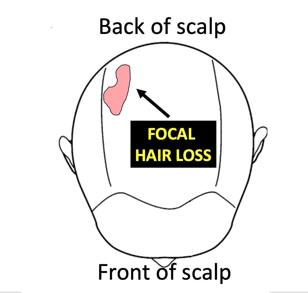
Focal alopecia refers to hair loss in a single area of the scalp. Common causes of focal alopecia include alopecia areata, tine capitis, infections, trauma, trichotillomania. Cancer is another cause of focal alopecia - albeit an uncommon one.

Scalp metastases refer to cancer the started in another organ and then the cancer spread to other parts of the body, including the scalp. Less than 2 % of patients that are known to have metastatic cancer will experience scalp metastases.


What are the most common cancers that metastasize to the scalp?

Lung cancer is the most common cancer that is associated with scalp metastases. Of all metastases to the scalp, lung cancer is the most common at 24 % followed by colon (11 %), liver (8 %) and breast (8%). Kidney and ovary remain other causes on the list. In 30 % of cases, the exact origin can’t be precisely determined. There are many types of ‘lung cancer’ and it remains debated as to which of the types is really the most likely contributor to scalp metastases (adenocarcinoma, large cell, small cell, squamous).


What are the clues that an area of hair loss may actually represent a metastasis?

Scalp metastases can be challenging to diagnose in the early stages. Sometimes they aren't large and sometimes they get overlooked. They may appear as papule or nodules, or firm indurated plaques or ulcers or as an area closely mimicking alopecia areata. They may simply appear as an area that looks like an infection. In many cases, they are red from dilated blood vessels - and sometimes hemorrhagic from localized blood clots in the area.  “Alopecia neoplastica” is a term that refers to hair loss associated with destruction of hair follicles. It is accompanied by red, indurated skin with dilated blood vessels and sometimes ulceration. In most cases, alopecia neoplastica represents hematologenous spread (spread in the blood stream) of a breast cancer. In some cases of metastastic scalp lesions, the areas of involvement are associated with no symptoms which in turn adds to the delay in diagnosis. In fact, there can be a delay in diagnosis for many patients with some studies showing a delay in proper diagnosis of 4–10 months following the time they are first noticed.

The identification of a scalp metastasis can rarely be the very first indication that the patient has a cancer inside the body. This is not common and in most cases it is already known that the patient has a cancer somewhere in the body. Overall, a study by Lookingbill and colleagues of 7316 cancer patients found that 0.8 % of patients had a skin lesion that represented the presenting sign of the cancer inside the body.

Key Conclusions and Summary

Scalp metastases can be challenging to diagnose in the early stages and diagnosis of a scalp metastasis ALWAYS comes from a biopsy. One can never look at a skin lesion and know with 100 % certainty that it is a metastasis from a cancer somewhere in the body. One only reaches that conclusion after a biopsy is done.

It’s often a good idea for a doctor to consider performing scalp biopsy when a patient has a focal areas of hair loss that does not improve over time - especially in patients over 45 years of age. In situations where the diagnosis is completely clea…

It’s often a good idea for a doctor to consider performing scalp biopsy when a patient has a focal areas of hair loss that does not improve over time - especially in patients over 45 years of age. In situations where the diagnosis is completely clear simply by examining the scalp, a biopsy is not necessary.


In general, a biopsy should at least be considered in the following situations:

1) A patient with a history of CANCER AT ANYTIME IN THE PAST who presents with a solitary area of hair loss (i.e. affecting a single localized area of the scalp).

2) A patient with CURRENT CANCER DIAGNOSIS who presents with a solitary area of hair loss (affecting a single localized area of the scalp).

3) A patient over 45 years of age who presents with a solitary LOCALIZED, RED PATCH OF HAIR LOSS ON THE SCALP that has persisted for 3 or more months.


Most patients with previous cancer diagnoses or who are dealing with cancer at the present time do not have a scalp metastasis as the reason for their hair problems when they present to the hair doctor’s office. However, it must always be on the doctor’s radar - especially when the patient has a solitary patch of hair loss (a single spot).

Persistent solitary patches of hair loss, especially when red, are extremely important to consider biopsying in anyone over 45.


Reference

Chiu CS, Lin CY, Kuo TT, et al: Malignant cutaneous tumors of the scalp: a study of demographic characteristics and histologic distributions of 398 Taiwanese patients. J Am Acad Dermatol. 56:448–452. 2007.

Frey L, Vetter-Kauczok C, Gesierich A, Bröcker EB and Ugurel S: Cutaneous metastases as the first clinical sign of metastatic gastric carcinoma. J Dtsch Dermatol Ges. 7:893–895. 2009

Kim HJ, Min HG and Lee ES: Alopecia neoplastica in a patient with gastric carcinoma. Br J Dermatol. 141:1122–1124. 1999.  

Lifshitz OH, Berlin JM, Taylor JS and Bergfeld WF: Metastatic gastric adenocarcinoma presenting as an enlarging plaque on the scalp. Cutis. 76:194–196. 2005

Lookingbill D.P., N. Spangler, F.M. SextonSkin involvement as the presenting sign of internal carcinoma. A retrospective study of 7316 cancer patients. J. Am. Acad. Dermatol., 22 (1) (1990), pp. 19-26


This article was written by Dr. Jeff Donovan, a Canadian and US board certified dermatologist specializing exclusively in hair loss.
Share This
No Comments

Permanent Chemotherapy Induced Alopecia (PCIA)

A New type of Hair loss in Patients Undergoing Chemotherapy: PCIA

Every year about 650,000 patients undergo chemotherapy in the United States. Hair loss is a common side effect of chemotherapy and occurs in about 65 % of patients who receive chemotherapy. There are two main types of hair loss that can occur in patients undergoing chemotherapy. The first is hair loss that happens within weeks of starting the chemotherapy and then lasts several months before growing back.  This is known as temporary chemotherapy induced alopecia ("TCIA"). The second type is uncommon and occurs when patients fail to regroth their hair back to the level it was before undergoing chemotherapy. If hair has not grown back after chemotherapy by the 6 month after chemotherapy, we call this permanent chemotherapy induced alopecia (PCIA) and it is sometimes also called Chemotherapy Induced Permanent Alopecia (CIPAL).


Permanent Chemotherapy Induced Alopecia (PCIA) 

The failure of the hair to grow back fully 6 months post chemotherapy raises concerns about a phenomenon known as permanent chemotherapy induced alopecia (PCIA).     In recent years a number of studies have highlighted the possibility of PCIA in women with breast cancer treated with various chemotherapeutic agents, especially drugs known as taxanes. Docetaxel and paclitaxel are part of this group of drugs. The exact mechanisms are unclear although injury to the bulb as well as follicular stem cells are thought to be relevant. Adjuvant anti-estrogen hormonal therapy may be an important cofactor in many women with PCIA. A similar PCIA presentation has been reported in patients undergoing bone marrow transplantation. The scalp is predominantly affected in women with PCIA although a minority may have eyebrow, eyelash and body hair loss as well.


Different Clinical Presentations of PCIA

PCIA doesn't appear similar in all patients. In fact, three main types appear to exist including a diffuse type, a diffuse type with vertex accentuation (mimicking androgenetic alopecia) and a patchy type mimicking alopecia areata.   


Examination under the Microscope: Biopsies of PCIA

Histopathology of biopsy specimens shows a non-scarring alopecia with preservation of sebaceous glands, miniaturization, decreased anagen hairs, increased telogen hairs and end stage avascular fibrous tracts. There may be several histological presentations and the exact features remains to be defined although a high proportion show dysmorphic telogen germinal units. Some biopsies show peribulbar type inflammation.


How do we treat PCIA?

We don't really know yet how to best treat PCIA. The most common treatments described in the medical literature are oral and topical minoxidil. Both seem to provide benefit to at least a proportion of patients.  Other treatments are not known to provide benefit. 


Dr. Donovan's Articles for Further Reading

Preventing Hair Loss from Chemotherapy

Does hair always grow back after chemotherapy?


REFERENCES

Miteva M, Misciali C, Fanti PA et al. Permanent alopecia after systemic chemotherapy: a clinicopathological study of 10 cases. Am J Dermatopathol. 2011 Jun;33(4):345-50.  

Fonia A, Cota C, Setterfield JF et al. Permanent alopecia in patients with breast cancer after taxane chemotherapy and adjuvant hormonal therapy: Clinicopathologic findings in a cohort of 10 patients. J Am Acad Dermatol. 2017 May;76(5):948-957.

Rugo HS.  Real-world use of scalp cooling to reduce chemotherapy-related hair loss.  Clin Adv Hematol Oncol. 2017


This article was written by Dr. Jeff Donovan, a Canadian and US board certified dermatologist specializing exclusively in hair loss.
Share This
No Comments

Does Elevated ESR cause Hair Loss?

The ESR Test

The erythrocyte sedimentation rate (ESR) is a very sensitive but non specific test for inflammation. An increased ESR does not directly cause hair loss but can sometimes indicate that the patient has underlying inflammation in the body that could be giving hair loss. Determining the cause of an elevated ESR is detective work.

eSR.png

The upper limit for ESR is slightly greater for women than men but a normal ESR is usually less than 20-30 mm/hr.

There are some conditions associated with a high ESR that are associated with hair loss and there are some conditions associated with high ESR that have nothing to do with hair loss. However, conditions such as various infections, and especially the autoimmune diseases (lupus, rheumatoid arthritis, vasculitis, inflammatory bowel disease), as well as anemias, pregnancy, some thyroid diseases, inflammatory diseases of the gastrointestinal tract and advanced kidney failure can be associated with hair loss. Other conditions including some cancers (especially blood cancers and various metastatic cancers) are associated with increased ESR but usually are not associated with hair loss. 

Very high ESR values over 100 mm/hr represent a special group. The group includes those that can be associated with hair loss include systemic lupus erythematosus, rheumatoid arthritis, and sometimes a few types of blood cancers (ie lymphomas, leukemias). Some drug hypersensitivity reactions can give very high ESR values and can also trigger hair loss. Polymyalgia rheumatica is in this group of conditions giving very high ESR values and can also sometimes give hair loss. Conditions in this group that usually don't give hair loss are infectious diseases such as abscesses, bacterial endocarditis and osteomyelitis.

The ESR test is a non specific test and many times a cause can't be found despite the patient having a full examination. Very high ESR levels may warrant additional testing. This may included other blood tests such as CRP, ANA, rheumatoid factor, LDH and possibly various imaging tests (depending on the precise history and precise level of ESR). There are hundreds of causes of increased ESR.


This article was written by Dr. Jeff Donovan, a Canadian and US board certified dermatologist specializing exclusively in hair loss.
Share This
1 Comment

Actinic Keratosis of the scalp

Scalp actinic keratoses

Actinic keratoses are red scaly lesions that develop from long term exposure to the sun. They are viewed as pre-cancers with the potential to become cancers known as squamous cell carcinomas (SCC).

Anywhere from 12 to 40% of individuals screened in dermatology clinics have actinic keratoses on the scalp. These lesions are far more common on the scalp in men than women, this is in part due to the increased exposure to ultraviolet radiation in men with hair loss. About 1-10 % of actinic keratoses will become a squamous cell carcinoma (SCC) over a 10 year period if left not treated. Approximately 1-2 % of SCC will metastasize making early treatment important.

References
Actinic keratosis: a cross-sectional study of disease characteristics and treatment patterns in Danish dermatology clinics. Erlendsson AM, et al. Int J Dermatol. 2016.

Prevalence of actinic keratosis among dermatology outpatients in Spain. Ferrándiz C, et al. Actas Dermosifiliogr. 2016.


This article was written by Dr. Jeff Donovan, a Canadian and US board certified dermatologist specializing exclusively in hair loss.
Share This
No Comments

Blogs by Topic


Share This
-->