h1.qusth1 { display: none !important; }

QUESTION OF THE WEEK

Dr. Donovan's Articles

QUESTION OF HAIR BLOGS


Pumpkin Seeds and Oil: What is the latest research?

What is the latest research?

PUMPKIN SEEDS.png

There’s little doubt that pumpkin seeds and pumpkin seed oil are good for most humans in modest amounts. However, there is still some debate as to whether pumpkin seeds and pumpkin seed oil (PSO) are beneficial for hair loss and how much exactly one should consume.

A good deal of excitement surfaced in 2014 when a study by Cho and colleagues showed that 40 % of men using 400 mg per day of pumpkin seed oil had measureable improvements in their “male balding” (androgenetic alopecia) compared to men in the same study using just placebo.

Surprisingly, good studies of pumpkin seed oil for treating hair loss have not been replicated nor published since that 2014 study first came out and so we are left with some uncertainty as to the true benefits of pumpkin seed oil.

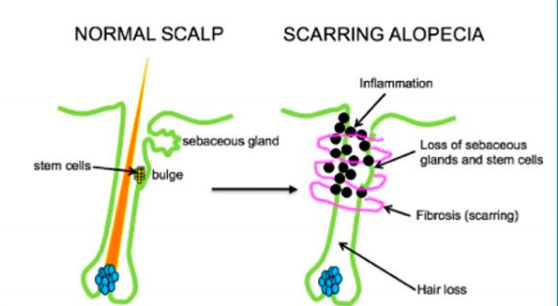
I continue to follow the medical research in the field. I’m interested not only in potential benefits for male balding but the potential for pumpkin seeds to affect the fats and lipids in the skin and of course the hair follicle. Many hair loss conditions, especially scarring alopecias, arise from altered “pro-inflammatory” lipids in the hair follicle. It’s clear that some dietary sources have a remarkable ability to change aspects of our physiology.

Pumpkin seeds are extremely rich in antioxidants and minerals. There is some evidence that they possess 5 alpha reductase abilities and so could impact conditions affected by androgens. Male balding, of course is the prototypical hair loss condition affected by androgens.

But pumpkin seed oil may have other anti-inflammatory properties. They may alter a variety of lipids but studies are limited to date. A recent study in rats showed that pumpkin seed oil at 100 mg/kg affected a variety of lipid metabolic enzymes such as fatty-acid synthase, acetyl CoA carboxylase, carnitine palmitoyl transferase-1, HMG CoA reductase. Pumpkin seed oil also reduced inflammatory markers such as TNF-α and IL-6.

Four years after the Cho research study, I don’t think we’re all that much further ahead in understanding the exact role of pumpkin seed oil when it comes to hair disorders. More research is needed. 


Reference

A K et al. Arch Physiol Biochem. 2018.


This article was written by Dr. Jeff Donovan, a Canadian and US board certified dermatologist specializing exclusively in hair loss.
Share This
No Comments

Scalp Pain (Trichodynia)

Dilated Blood Vessels Frequently Seen in Trichodynia

trichodynia

“Trichodynia” is a somewhat poorly defined term that generally refers to hair pain. To learn what trichodynia really feels like, I encourage physicians I teach to wear a hat (especially a heavy helmet) for many hours and then feel what it’s like when one removes the hat and proceeds to move their hair from one side to the other. The unpleasant experience is ... trichodynia! (and it hurts!). There are many potential causes of trichodynia and so anyone with these symptoms needs careful evaluation. Scarring alopecias, alopecoa areata, telogen effluvium, stress, anxiety, depression all can cause trichodynia.

A study by Willimann and Trueb (in 2002) showed that dilated blood vessels seen in the scalp were strongly associated with trichodynia. The reasons still are not entirely clear but may be related to the release of the neuropeptide known as “substance P.” Substance P is a potent vasodilator and may explain (at least partly) the blood vessel dilation sometimes observed in patients with trichodynia.


REFERENCE

Willimann B, et al. Hair pain (trichodynia): frequency and relationship to hair loss and patient gender. Dermatology. 2002.


This article was written by Dr. Jeff Donovan, a Canadian and US board certified dermatologist specializing exclusively in hair loss.
Share This
9 Comments

Steroid Injections: Helpful for Inflammatory Hair Loss Conditions

Helpful for Inflammatory Hair Loss Conditions

STEROID INJ.png

"Steroid injections" (using triamcinolone acetonide) are helpful for a variety of inflammatory hair loss conditions including alopecia areata, lichen planopilaris, frontal fibrosing alopecia, pseudopelade, discoid lupus, central centrifugal cicatricial alopecia, psoriasis and others.

Steroid injections are one of the most consistently helpful treatments to combat problematic scalp inflammation. When used at the right dose (2.5 to 10 mg/mL for 20 mg max) and at the right frequency (every 5-12 weeks) they have a reasonably good safety profile.

Steroid injections can have a very important role in a well thought out treatment plan for many inflammatory conditions.


This article was written by Dr. Jeff Donovan, a Canadian and US board certified dermatologist specializing exclusively in hair loss.
Share This
No Comments

Hydroxychloroquine (Plaquenil): How does it work?

How does it work?

Plaquenil.png

Hydroxychloroquine (HCQ) is an oral medication widely used for many types of inflammatory and autoimmune type conditions. "Plaquenil" is a popular trade name but there are many generics available. In the world of hair loss, these drugs are commonly used to treat scarring alopecias such as lichen planopilaris, frontal fibrosing alopecia, discoid lupus and pseudopelade.

How does the drug work? Hydroxychloroquine is mildly immunosuppressive. The precise mechanism of action has not been entirely elucidated but dozens of different mechanisms have been uncovered.

HCQ has an unusual property in that it accumulates in a specific area of cells known as “lysosomes” and “endosomes.” By doing so, it changes the pH in these compartments and interferes with the normal jobs that are supposed to go on inside these cellular compartments. Many cells of the immune system, including macrophages, lymphocytes and neutrophils, rely on their lysosomes to function properly.

As a result, HCQ interferes with a range of normal processes of the immune system including interfering with antigen processing.  They are known to 1) inhibit cytokine release of key molecules that activate the immune system (IL) 1, IL-2, IL-6, IL-18, tumor necrosis factor α, interferon γ) 2) decrease the activity of NK cells 3) affect neutrophil function 4) regulate apoptosis and 5) inhibit the activity of T cells.


This article was written by Dr. Jeff Donovan, a Canadian and US board certified dermatologist specializing exclusively in hair loss.
Share This
No Comments

Do patients with LPP have an increased risk of thyroid disease?

Thyroid Disease Present in 1 out of 3 Patients with Lichen Planopilaris


A variety of blood tests are important for all patients with hair loss. The exact tests that are needed vary somewhat depending on the specific type of hair loss in question. For patients with lichen planopilaris, thyroid studies must always be part of a general work-up. 

In 2014, Mesinkovska and colleagues at the Cleveland Clinic set out to evaluate the frequency of thyroid disorders in patients with lichen planopilaris. They evaluated the medical records of 166 patients with LPP. They compared the thyroid lab work in these patients to 81 age- and gender-matched control subjects.

Interestingly, a diagnosis of thyroid disease was present in 34% (n = 57) of the 166 patients with LPP, but just 11% (n = 9) of the control subjects. When confined to hypothyroidism alone, this disease was found in 29% (n = 48) of the patients with LPP and 9% (n = 7) of the control subjects.


Conclusion

Hyperthyroidism is common in lichen planopilaris. This data suggests that about 1 in every 3 patients may have thyroid disease. Screening tests for thyroid abnormalities are essential in all patients with lichen planopilaris.


Reference

Atanaskova Mesinkovska N, et al. Association of lichen planopilaris with thyroid disease: a retrospective case-control study. J Am Acad Dermatol. 2014.


This article was written by Dr. Jeff Donovan, a Canadian and US board certified dermatologist specializing exclusively in hair loss.
Share This
No Comments

Yellow dots in Androgenetic Alopecia

Yellow Dots Represent Sebaceous Material

YD

"Yellow dots" can be seen in many hair loss conditions including alopecia areata and androgenetic alopecia. In androgenetic alopecia, the yellow dots represent hair follicle openings packed with sebum and keratin material. The yellow dots can be easily seen if a patient has not shampooed the scalp for a few days. However, the disappear with washing. In contrast to the yellow dots of alopecia areata, the yellow dots seen in AGA are much more variable in size.


This article was written by Dr. Jeff Donovan, a Canadian and US board certified dermatologist specializing exclusively in hair loss.
Share This
No Comments

Knowing when not to prescribe

To prescribe or not to prescribe

Knowing when not to prescribe a medication is just as important as knowing when to prescribe it.

prescribing


Should be 24 year old male with male balding and severe depression be prescribed finasteride? What about the the 57 year old male with male pattern balding and unstable angina. Can he use minoxidil?  Should the 31 year old female with folliculitis decalvans on isotretinoin also receive doxycycline? Should the 45 year old female with lichen planopilaris and pre-existing retinopathy receive hydroxychloroquine?  The answer to all of these questions is no.

Knowing when not to prescribe a medication is just as important as knowing when to prescribe it.


This article was written by Dr. Jeff Donovan, a Canadian and US board certified dermatologist specializing exclusively in hair loss.
Share This
No Comments

Can hydroxychloroquine (Plaquenil) be prescribed in the setting of G6PD deficiency

Hydroxychloroquine in G6PD Deficiency

Hydroxychloroquine (Plaquenil) is an oral immunomodulating medication commonly used for the the treatment of autoimmune diseases. In the world of hair loss, it is commonly used for treating lichen planopilaris, frontal fibrosing alopecia, discoid lupus and pseudopelade. Some reports have emerged that it may even be useful in alopecia areata. 

For years, physicians have been taught that certain groups of patients should not be prescribed hydroxychloroquine. These include patients with psoriasis, retinal problems, certain psychiatric disorders, porphyria, anemias, neutropenias, and liver problems. In addition, patients with deficiency of an enzyme known as Glucose-6-Phosphate Dehydrogenase (G6PD) were also thought to be ineligible for the mediation given their increased risk of hemolytic anemia. 

About 400 million people have G6PD deficiency. It is more common in the Middle East, Mediterranean, and parts of Africa and Asia.  It is a genetic condition that is present from birth. Without the enzyme, patients experience hemolysis of death of their own red blood cells from triggers like infection, medications, stress and even some foods (i.e. fava beans). The actual severity of the condition varies greatly depending on the specific enzyme that is inherited at birth.

New Studies suggest Plaquenil may be safer in G6PD deficiency than once thought

I was very interested to recently read an abstract by Samya Mohammad and colleagues at Duke University presented at the 2016 meeting of the American College of Rheumatology. The abstract was titled " Hydroxychloroquine Is Not Associated with Hemolytic Anemia in Glucose-6-Phosphate Dehydrogenase (G6PD) Deficient Patients" 

The authors set out to evaluate 275 patients who were prescribed hydroxychloroquine including 11 who were G6PD deficient.   The study did not involve hair patients but rather patients with diagnoses such as lupus (32%), rheumatoid arthritis (29%), and other inflammatory joint problems (14%).  

The G6PD deficient patients had a total of 711 months of exposure to ydroxychloroquine.  In this cohort, no G6PD deficient patients developed hemolytic anemia attributable to the drug during 711 months exposure to the drug.


Conclusion

To date this remains the largest study to date evaluating the frequency of hemolytic anemia in  G6PD deficient patients treated with hydroxychloroquine. Although small in number, the authors felt that their data do not support routine G6PD level measurement prior to initiating HCQ therapy.

Reference

Mohammad S, Clowse MEB, Eudy A, Criscione-Schreiber L. Hydroxychloroquine Is Not Associated with Hemolytic Anemia in Glucose-6-Phosphate Dehydrogenase (G6PD) Deficient Patients [abstract]. Arthritis Rheumatol. 2016; 68 (suppl 10). https://acrabstracts.org/abstract/hydroxychloroquine-is-not-associated-with-hemolytic-anemia-in-glucose-6-phosphate-dehydrogenase-g6pd-deficient-patients/. Accessed June 23, 2018.


This article was written by Dr. Jeff Donovan, a Canadian and US board certified dermatologist specializing exclusively in hair loss.
Share This
No Comments

Focal atrichia in androgenetic alopecia

What is the significance of focal atrichia?

FA-AGA

"Focal atrichia" (FA) refers to the absence of hairs is defined areas on the scalp. It is a feature generally seen in androgenetic alopecia whereby small circular areas totally devoid of hair are seen. These areas are typically the size of a pencil eraser and sometimes a bit bigger. Biopsies of these ares in the setting of androgenetic alopecia show accumulation of tiny vellus hairs.


It’s clear that FA is more common in androgenetic alopecia than other types of hair loss. A 2017 study by Olsen and colleagues showed FA was seen in 44 % of patients with FPHL compared to 2 % of other diseases. Those other 2 % may include area of primary scarring alopecia, scars from trauma, and even diffuse alopecia areata. Nevertheless, focal atrichia is most commonly seen in androgenetic alopecia.

There are still a few things that are unclear about whether one sees FA atrichia only in advanced disease. That same 2017 study by Olsen suggested 67 % of women with “late onset” FPHL had FA compared to just 15 % of those with “early onset” AGA. As we continue our discussion one must keep in mind that late onset generally has better prognosis.


Study 1: Olsen and colleagues, 2017

The 2017 Olsen study set out to evaluate the frequency of focal atrichia in various types of hair loss and its histologic characteristics in female androgenetic alopecia. The authors reviewed 250 consecutive female patients seen with hair loss for the presence or absence of FA. Interestingly, FA was identified in 46/104 (44%) of women with female pattern hair loss, including 15 % of early onset and 67% of late onset compared to 3/146 (2%) of those with other hair disorders. The histological (biopsy) findings of FA in FPHL showed mainly a more progressive miniaturization process than that of haired areas of the scalp. Taken together Olsen and her colleagues concluded that FA wasa clinical clue to the diagnosis of FPHL, particularly late onset subtype.


Study 2: Hu and colleagues, 2015

Studies by both Hu and colleagues and Zhang and colleagues both independently showed FA was associated with more severe disease. Additionally, Zhang et al showed that it was also associated with longer duration of disease. These findings seem to be at odds with the Olsen study.

Hu and colleagues performed a case-control observational study to identify the trichoscopic findings of AGA and to evaluate their relationship with the overall severity of the androgenetic aloepcia. The authors performed trichoscopic examination for 750 male AGA patients and 200 female (FPHL) patients, along with 100 male and 50 female normal controls.  In this study, FA was positively related to severity of hair loss (P < 0.05). 


Study 3: Hu and colleagues, 2012

Zhang and colleagues set out to analyze characteristics and investigate associations and clinical and trichoscopic features of female patients with FPHL. The did so by evaluating data from 60 patients with FPHL. FA was positively correlated with the stage and duration of hair loss.
 

Taken together, it is clear that FA is frequently seen in androgenetic alopecia. At first glance, is not uncommon to wonder whether these areas in fact represent scarring alopecia (i.e. tiny patches of Pseudopelade of Brocq or lichen planopilaris) or represent small areas of alopecia areata. However, when taken in context with the miniaturization going on in other areas of the scalp nearby, one can generally be confident these are area of FA. 

The general consensus would be the FA is a negative prognostic factor. 


Reference

Olsen et al. Focal Atrichia: A Diagnostic Clue in Female Pattern Hair Loss.
J Am Acad Dermatol. 2017.

Hu R, et al. Trichoscopic findings of androgenetic alopecia and their association with disease severity. J Dermatol. 2015.

Zhang X, et al. Female pattern hair loss: clinico-laboratory findings and trichoscopy depending on disease severity.  Int J Trichology. 2012.


This article was written by Dr. Jeff Donovan, a Canadian and US board certified dermatologist specializing exclusively in hair loss.
Share This
No Comments

Fusion Follicles: Compound Follicles = More than 6 Hairs

Compound Follicles = More than 6 Hairs

compound follicles 6 hairs.png

Compound Hair Follicles are hair follicles with more than the normal number of hairs emerging from a single hair follicle opening. In this photo, we see some of the earliest signs of a condition known as folliculitis decalvans. The scalp is red and scaly and there is a clear tendency for hair follicles to fuse together. The arrow points to 9 hair follicles all grouped together and emerging from the single opening. This so called compound follicle is commonly seen in many patients with folliculitis decalvans as well as some other scarring alopecias as well.

Treatment with topical antibiotics, oral antibiotics, retinoids, topical and/or intralesional corticosteroids are the best primary options in managing the disease.


This article was written by Dr. Jeff Donovan, a Canadian and US board certified dermatologist specializing exclusively in hair loss.
Share This
No Comments

The Scalp Punch Biopsy: Will anyone notice I had it done?

scalp bx- noticable?.png

Will anyone notice I had it done?

A punch biopsy of the scalp is performed if their is any uncertainty about the diagnosis of a patient’s hair loss. It involves removal of a 4 mm cylindrical core of tissue and placement of a stitch (suture). A punch biopsy should be taken from an area showing the main features of the disease in question. Provided the biopsy is taken from an area that contains a reasonable amount of hair, the stitch and healing site should be relatively unnoticeable. A dissolving suture or non dissolving suture can be placed although I tend to prefer dissolving sutures.


This article was written by Dr. Jeff Donovan, a Canadian and US board certified dermatologist specializing exclusively in hair loss.
Share This
No Comments

Scarring Alopecia: Is it inactive?

Is it inactive?

SA-inactive?.png

There are many misconceptions when it comes to determining if a scarring alopecia is truly “quiet.” Scarring alopecias, are a group of hair conditions whereby the body forms scar tissue in the scalp. This scar tissue ultimately prevents hairs from growing properly.

Here are a few principles that should always be considered when one feels they want to say a scarring alopecia is “quiet” or “inactive” 1) No doctor can tell if a scarring alopecia is quiet by looking at the scalp on a first appointment visit. That sentence deserves reading twice. Some scarring alopecias look quiet but the patient continues to lose hair. You can really only say a scarring alopecia is quiet if you’ve re-examined the scalp 12-24 months after the first appointment. 2) A patient with scalp symptoms like itching, burning or pain probably does not have quiet (inactive) disease. 3) Patients with redness and scaling in the scalp probably have active disease but they may not. Redness can be a sign of active disease but some patients with prolonged use of topical steroids simply have a red scalp. 4) The most important (and too often forgotten) guide that determines if a scarring alopecia is “quiet” is a comparison of scalp photographs taken at two different time points. Provided the interval between photos is greater than 1 year, one can get some pretty good information about whether the condition is quiet.

In my opinion, if there is no change in hair density between 2 photographs taken ONE year apart, the scarring alopecia is “probably” quiet (inactive). If there is no change in hair density between 2 photographs taken TWO years apart, the scarring alopecia is “extremely likely” to be deemed quiet. Despite this inactive appearance, the scarring alopecia STILL carries a risk of reactivation.

If there is no change in hair density between 2 photographs taken THREE years apart, and the patient is OFF MEDICATION during that period of observation, the scarring alopecia is likely burnt out.


This article was written by Dr. Jeff Donovan, a Canadian and US board certified dermatologist specializing exclusively in hair loss.
Share This
2 Comments

Alopecia Areata

Alopecia Areata: A Cause of Non Scarring Hair Loss

AA?.png

Alopecia areata is a form of autoimmune hair loss. Inflammation occuring deep under the scalp causes hair to fall out. The hair loss is non scarring which means the potential exists for hair to grow back. 


This photo shows the scalp of a man with advanced alopecia areata. Yellow dots represent keratin plugged openings of the hair follicles. Despite his widespead hair loss (involving 98% of this patient’s scalp), there are hairs on the scalp that appear completely unaffected by the immune reaction. Why that is remains unknown. Treatments for advanced alopecia areata include steroid injections, diphencyprone, anthralin, methotrexate, sulfasalazine, hydroxycloroquine and tofacitinib 


This article was written by Dr. Jeff Donovan, a Canadian and US board certified dermatologist specializing exclusively in hair loss.
Share This
2 Comments

Lichen Planopilaris: Itching, Burning, Tenderness, Shedding

Itching, Burning, Tenderness, Shedding

LPP symptoms.png

Lichen planopilaris (LPP) is a scarring hair loss condition that leads to permanent hair loss in affected areas without treatment.

Individuals with the condition may initially develop itching, burning or pain. For some, increased amounts of shedding may be the only symptom.

Examination of LPP via up close (“dermatoscopic”) examination is shown here. The scalp is red and scaly. In particular, some of the scale is located around hair follicles (called perifollicular scale). There are distinct whitish areas seen which represent permanently scarred areas of the scalp.

Treatment of LPP includes topical steroids, topical calcineurin inhibitors, steroid injections and a variety of oral immunosuppressive medications such as doxycycline, hydroxychloroquine, mycophenolate, cyclosporine, methotrexate, isotretinoin, tofacitinib and pioglitazone. Low level laser and excimer laser could to be studied and appear to offer benefit for some.


This article was written by Dr. Jeff Donovan, a Canadian and US board certified dermatologist specializing exclusively in hair loss.
Share This
No Comments

Corticosteroid Telangiectasias

Corticosteroid Telangiectasias

corticosteroids.png

Topical steroids have an important role in the treatment of hair loss. Like any medication, they must be respected. 


Dilatation of blood vessels (telangiectasias) can occur with prolonged topical steroid use. This is thought to occur due to stimulation of release of a chemical known as nitric oxide (NO) from dermal vessel endothelial cells which in turn leads to abnormal blood vessel dilatation. This photo shows telangiectasias occurring in a patient with alopecia areata treated with prolonged topical clobetasol. 


This article was written by Dr. Jeff Donovan, a Canadian and US board certified dermatologist specializing exclusively in hair loss.
Share This
2 Comments

Scarring Alopecia: Top 25 Frequently Asked Questions

SA 25 questions.png

Top 25 Frequently Asked Questions

Scarring alopecia (also called cicatricial alopecia) is a form of hair loss that has the potential to cause permanent hair loss. 

These conditions are not as common as hereditary balding which gives rise to many myths, misunderstandings and miscommunications when it comes to the diagnosis and treatment of these conditions.

Our patient handout can be downloaded here

PATIENT INFORMATION - TOP 25 FAQ SCARRING ALOPECIA


This article was written by Dr. Jeff Donovan, a Canadian and US board certified dermatologist specializing exclusively in hair loss.
Share This
2 Comments

What is scarring alopecia?

scarrinf A.png

What are scarring alopecias?

Scarring alopecias (also referred to as the cicatricial alopecias) are a broad group of hair loss conditions that are associated with inflammation and scarring. Inflammation in the upper parts of the hair follicle leads to destruction of hair loss stem cells and loss of sebaceous glands.

The slow development of fibrosis (scarring) leads to a permanent loss of hairs.


This article was written by Dr. Jeff Donovan, a Canadian and US board certified dermatologist specializing exclusively in hair loss.
Share This
No Comments

Caffeine and Hair: What does Caffeine do for Hair?

What does caffeine do for hair?

caffeone.png

This specialty coffee reminded me about the increasing attention recently on topical (not oral) caffeine for hair growth. Topical caffeine is slowly catching the attention of the hair world and general public. A small number of studies have suggested that caffeine may have positive effects on hair follicles when applied topically. Some shampoos are even including caffeine additives with “claims” it reduces shedding.

I’m reminded of a 2014 study which set out to investigate the impact of caffeine on hair growth in vitro and to better understand how caffeine modulates the effects of testosterone.

The researchers used microdissected human scalp hair follicles (both male and female) and treated them in culture with testosterone or the combination of testosterone and caffeine). Remarkably, caffeine prolonged anagen duration, enhanced hair shaft elongation and stimulated hair matrix keratinocyte proliferation. Hair follicles from men were less sensitive to caffeine than female follicles.

Caffeine had effects on TGF-β2 and IGF-1. Specifically, caffeine blocked testosterone-enhanced TGF-β2 protein expression in male HFs. In female HFs, caffeine reduced TGF-β2 expression. IGF-1 protein expression was upregulated in both male and female hair follicles.

I have read and reread this study a few times over the years. It’s quite interesting that caffeine has hair growth promoting properties.

The use topical caffeine for its effects on hair is still in its early days. There are several commercially available products that contain caffeine (mainly shampoos) and a few of these shampoos have small clinical studies that show some potential to reduce shedding and improve the look of the hair. These studies are small and preliminary in nature.

Government regulators in some countries have recently come down hard on manufacturers of “caffeine” containing hair products (especially some shampoo makers) scrutinizing claims that these products benefit hair. More rigorous studies are needed to determine definitively what role these products have.
 

Reference

Fischer TW, et al. Br J Dermatol. 2014. and Sisto et al J Appl Cosmetol 2013


This article was written by Dr. Jeff Donovan, a Canadian and US board certified dermatologist specializing exclusively in hair loss.
Share This
No Comments

Alopecia areata and Androgens: Are we missing something?

AA and Androgens

Alopecia areata is an autoimmune disease. It occurs in about 2 % of the population and in all age groups and races. To date the role of hormones in alopecia areata is unknown – although the area is poorly studied in general.

In 2017, Conic and colleagues set out to retrospectively review the clinical features of patients with alopecia areata that were seen at the Cleveland Clinic over the period 2005 to 2014.  In total, data from 504 patients was tabulated and as a comparison group 172 patients with seborrheic dermatitis were also reviewed. 

Elevated androgens were far more common in those with alopecia areata compared to controls (13 % vs 1 %).  Ovarian cysts were also more frequent in those with AA being present in 8.6 % of patients vs just 3.2 % of controls. 


Conclusion

This study was interesting and certainly caught my attention. Very little is known about how hormones affect AA. The increase incidence of androgens warrants further study. If consistently found in a proportion of patients, one needs to explore whether use of anti-androgens could benefit some patients with AA as well.  In the same light, one needs to consider whether androgenic progestins in oral contraceptives could act as a trigger for AA in some patients


REFERENCE

Conic et al. Comorbidities in patients with alopecia areata. Journal American Academy Dermatology; 754-756.


This article was written by Dr. Jeff Donovan, a Canadian and US board certified dermatologist specializing exclusively in hair loss.
Share This
No Comments

Deficiencies of iron and vitamin D in patients with alopecia areata

Deficiencies of iron and vitamin D in patients with alopecia areata

Alopecia areata is an autoimmune disease. Blood tests are important for patients with alopecia areata given that recent research has suggested that vitamin D deficiency as well as other deficiencies such as iron deficiency may be more common in alopecia areata.  

In 2017, Conic and colleagues set out to retrospectively review the clinical features of patients with alopecia areata that were seen at the Cleveland Clinic over the period 2005 to 2014.  In total, data from 504 patients was tabulated and as a comparison group 172 patients with seborrheic dermatitis were also reviewed. 

Patients with alopecia areata had more frequent vitamin D deficiency (30 % compared to 13 % in controls) and also had more frequent iron deficiency (7.3 % vs 2.9 % of controls).  Anemia was also more common being present in 17 % of those with alopecia areata and only 7.6% of control patients. 


Conclusion

Deficiencies of iron and vitamin D are more common in alopecia areata. Testing levels of iron nd vitamin D are important in alopecia areata.


REFERENCE

Conic et al. Comorbidities in patients with alopecia areata. Journal American Academy Dermatology; 754-756.


This article was written by Dr. Jeff Donovan, a Canadian and US board certified dermatologist specializing exclusively in hair loss.
Share This
No Comments


Share This
-->